Inung Wijayanto, Rudy Hartanto, Hanung Adi Nugroho
{"title":"癫痫脑电图信号的半球间和半球内相干性定量分析。","authors":"Inung Wijayanto, Rudy Hartanto, Hanung Adi Nugroho","doi":"10.4103/jmss.JMSS_63_20","DOIUrl":null,"url":null,"abstract":"<p><p>When an epileptic seizure occurs, the neuron's activity of the brain is dynamically changed, which affects the connectivity between brain regions. The connectivity of each brain region can be quantified by electroencephalography (EEG) coherence, which measures the statistical correlation between electrodes spatially separated on the scalp. Previous studies conducted a coherence analysis of all EEG electrodes covering all parts of the brain. However, in an epileptic condition, seizures occur in a specific region of the brain then spreading to other areas. Therefore, this study applies an energy-based channel selection process to determine the coherence analysis in the most active brain regions during the seizure. This paper presents a quantitative analysis of inter- and intrahemispheric coherence in epileptic EEG signals and the correlation with the channel activity to glean insights about brain area connectivity changes during epileptic seizures. The EEG signals are obtained from ten patients' data from the CHB-MIT dataset. Pair-wise electrode spectral coherence is calculated in the full band and five sub-bands of EEG signals. The channel activity level is determined by calculating the energy of each channel in all patients. The EEG coherence observation in the preictal (<i>Coh<sub>pre</sub></i> ) and ictal (<i>Coh<sub>ictal</sub></i> ) conditions showed a significant decrease of <i>Coh<sub>ictal</sub></i> in the most active channel, especially in the lower EEG sub-bands. This finding indicates that there is a strong correlation between the decrease of mean spectral coherence and channel activity. The decrease of coherence in epileptic conditions (<i>Coh<sub>ictal</sub></i> <<i>Coh<sub>pre</sub></i> ) indicates low neuronal connectivity. There are some exceptions in some channel pairs, but a constant pattern is found in the high activity channel. This shows a strong correlation between the decrease of coherence and the channel activity. The finding in this study demonstrates that the neuronal connectivity of epileptic EEG signals is suitable to be analyzed in the more active brain regions.</p>","PeriodicalId":37680,"journal":{"name":"Journal of Medical Signals & Sensors","volume":"12 2","pages":"145-154"},"PeriodicalIF":1.1000,"publicationDate":"2022-05-12","publicationTypes":"Journal Article","fieldsOfStudy":null,"isOpenAccess":false,"openAccessPdf":"https://ftp.ncbi.nlm.nih.gov/pub/pmc/oa_pdf/da/54/JMSS-12-145.PMC9215829.pdf","citationCount":"0","resultStr":"{\"title\":\"Quantitative Analysis of Inter- and Intrahemispheric Coherence on Epileptic Electroencephalography Signal.\",\"authors\":\"Inung Wijayanto, Rudy Hartanto, Hanung Adi Nugroho\",\"doi\":\"10.4103/jmss.JMSS_63_20\",\"DOIUrl\":null,\"url\":null,\"abstract\":\"<p><p>When an epileptic seizure occurs, the neuron's activity of the brain is dynamically changed, which affects the connectivity between brain regions. The connectivity of each brain region can be quantified by electroencephalography (EEG) coherence, which measures the statistical correlation between electrodes spatially separated on the scalp. Previous studies conducted a coherence analysis of all EEG electrodes covering all parts of the brain. However, in an epileptic condition, seizures occur in a specific region of the brain then spreading to other areas. Therefore, this study applies an energy-based channel selection process to determine the coherence analysis in the most active brain regions during the seizure. This paper presents a quantitative analysis of inter- and intrahemispheric coherence in epileptic EEG signals and the correlation with the channel activity to glean insights about brain area connectivity changes during epileptic seizures. The EEG signals are obtained from ten patients' data from the CHB-MIT dataset. Pair-wise electrode spectral coherence is calculated in the full band and five sub-bands of EEG signals. The channel activity level is determined by calculating the energy of each channel in all patients. The EEG coherence observation in the preictal (<i>Coh<sub>pre</sub></i> ) and ictal (<i>Coh<sub>ictal</sub></i> ) conditions showed a significant decrease of <i>Coh<sub>ictal</sub></i> in the most active channel, especially in the lower EEG sub-bands. This finding indicates that there is a strong correlation between the decrease of mean spectral coherence and channel activity. The decrease of coherence in epileptic conditions (<i>Coh<sub>ictal</sub></i> <<i>Coh<sub>pre</sub></i> ) indicates low neuronal connectivity. There are some exceptions in some channel pairs, but a constant pattern is found in the high activity channel. This shows a strong correlation between the decrease of coherence and the channel activity. The finding in this study demonstrates that the neuronal connectivity of epileptic EEG signals is suitable to be analyzed in the more active brain regions.</p>\",\"PeriodicalId\":37680,\"journal\":{\"name\":\"Journal of Medical Signals & Sensors\",\"volume\":\"12 2\",\"pages\":\"145-154\"},\"PeriodicalIF\":1.1000,\"publicationDate\":\"2022-05-12\",\"publicationTypes\":\"Journal Article\",\"fieldsOfStudy\":null,\"isOpenAccess\":false,\"openAccessPdf\":\"https://ftp.ncbi.nlm.nih.gov/pub/pmc/oa_pdf/da/54/JMSS-12-145.PMC9215829.pdf\",\"citationCount\":\"0\",\"resultStr\":null,\"platform\":\"Semanticscholar\",\"paperid\":null,\"PeriodicalName\":\"Journal of Medical Signals & Sensors\",\"FirstCategoryId\":\"1085\",\"ListUrlMain\":\"https://doi.org/10.4103/jmss.JMSS_63_20\",\"RegionNum\":0,\"RegionCategory\":null,\"ArticlePicture\":[],\"TitleCN\":null,\"AbstractTextCN\":null,\"PMCID\":null,\"EPubDate\":\"2022/4/1 0:00:00\",\"PubModel\":\"eCollection\",\"JCR\":\"Q4\",\"JCRName\":\"ENGINEERING, BIOMEDICAL\",\"Score\":null,\"Total\":0}","platform":"Semanticscholar","paperid":null,"PeriodicalName":"Journal of Medical Signals & Sensors","FirstCategoryId":"1085","ListUrlMain":"https://doi.org/10.4103/jmss.JMSS_63_20","RegionNum":0,"RegionCategory":null,"ArticlePicture":[],"TitleCN":null,"AbstractTextCN":null,"PMCID":null,"EPubDate":"2022/4/1 0:00:00","PubModel":"eCollection","JCR":"Q4","JCRName":"ENGINEERING, BIOMEDICAL","Score":null,"Total":0}
Quantitative Analysis of Inter- and Intrahemispheric Coherence on Epileptic Electroencephalography Signal.
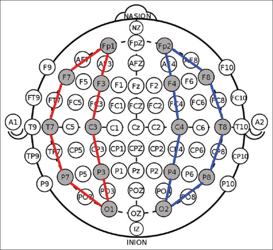
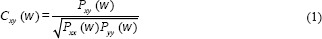
When an epileptic seizure occurs, the neuron's activity of the brain is dynamically changed, which affects the connectivity between brain regions. The connectivity of each brain region can be quantified by electroencephalography (EEG) coherence, which measures the statistical correlation between electrodes spatially separated on the scalp. Previous studies conducted a coherence analysis of all EEG electrodes covering all parts of the brain. However, in an epileptic condition, seizures occur in a specific region of the brain then spreading to other areas. Therefore, this study applies an energy-based channel selection process to determine the coherence analysis in the most active brain regions during the seizure. This paper presents a quantitative analysis of inter- and intrahemispheric coherence in epileptic EEG signals and the correlation with the channel activity to glean insights about brain area connectivity changes during epileptic seizures. The EEG signals are obtained from ten patients' data from the CHB-MIT dataset. Pair-wise electrode spectral coherence is calculated in the full band and five sub-bands of EEG signals. The channel activity level is determined by calculating the energy of each channel in all patients. The EEG coherence observation in the preictal (Cohpre ) and ictal (Cohictal ) conditions showed a significant decrease of Cohictal in the most active channel, especially in the lower EEG sub-bands. This finding indicates that there is a strong correlation between the decrease of mean spectral coherence and channel activity. The decrease of coherence in epileptic conditions (Cohictal <Cohpre ) indicates low neuronal connectivity. There are some exceptions in some channel pairs, but a constant pattern is found in the high activity channel. This shows a strong correlation between the decrease of coherence and the channel activity. The finding in this study demonstrates that the neuronal connectivity of epileptic EEG signals is suitable to be analyzed in the more active brain regions.
期刊介绍:
JMSS is an interdisciplinary journal that incorporates all aspects of the biomedical engineering including bioelectrics, bioinformatics, medical physics, health technology assessment, etc. Subject areas covered by the journal include: - Bioelectric: Bioinstruments Biosensors Modeling Biomedical signal processing Medical image analysis and processing Medical imaging devices Control of biological systems Neuromuscular systems Cognitive sciences Telemedicine Robotic Medical ultrasonography Bioelectromagnetics Electrophysiology Cell tracking - Bioinformatics and medical informatics: Analysis of biological data Data mining Stochastic modeling Computational genomics Artificial intelligence & fuzzy Applications Medical softwares Bioalgorithms Electronic health - Biophysics and medical physics: Computed tomography Radiation therapy Laser therapy - Education in biomedical engineering - Health technology assessment - Standard in biomedical engineering.


 求助内容:
求助内容: 应助结果提醒方式:


