{"title":"肾小管囊性细胞癌:一个未被充分认识的临床病理实体。","authors":"Sudeep Khera, Poonam Elhence, Taruna Yadav, Himanshu Pandey","doi":"10.31486/toj.21.0065","DOIUrl":null,"url":null,"abstract":"<p><p><b>Background:</b> Tubulocystic renal cell carcinoma is a lesser-known neoplastic entity compared to other common histologic variants of renal cell carcinoma. The World Health Organization identified tubulocystic renal cell carcinoma as a newly recognized renal tumor in 2016. We report a case of tubulocystic renal cell carcinoma in a young adult. <b>Case Report:</b> A 21-year-old male presented with the chief complaint of a lump on the right side of his abdomen since childhood. Magnetic resonance imaging revealed an enlarged right kidney with multiple large multiloculated cysts with hemorrhagic contents and enhancing peripheral nodular solid components with enhancing septa in some of the cysts, suggesting the possibility of multifocal intracystic papillary renal cell carcinoma. Imaging showed a Bosniak type IV cystic lesion in the right kidney. Right radical nephrectomy was performed. Grossly, the kidney was almost replaced by variable-sized cystic lesions with thick septations filled with serous fluid to gelatinous material. The tubules and cysts were lined by a single layer of flat, hobnail, and cuboidal cells with high-grade nuclear features. No ovarian-type stroma was identified. In places, a papillary component was also identified. Tubulocystic renal cell carcinoma was diagnosed based on microscopy and immunohistochemistry results. <b>Conclusion:</b> Tubulocystic renal cell carcinoma is a rare entity that was previously called Bellini duct carcinoma and low-grade collecting duct carcinoma. Because of the limited number of cases reported, tubulocystic renal cell carcinoma needs to be followed to determine the biologic behavior and metastatic potential of these tumors so that management guidelines for such cases can be developed.</p>","PeriodicalId":47600,"journal":{"name":"Ochsner Journal","volume":"22 2","pages":"182-187"},"PeriodicalIF":1.3000,"publicationDate":"2022-01-01","publicationTypes":"Journal Article","fieldsOfStudy":null,"isOpenAccess":false,"openAccessPdf":"https://www.ncbi.nlm.nih.gov/pmc/articles/PMC9196962/pdf/","citationCount":"0","resultStr":"{\"title\":\"Tubulocystic Renal Cell Carcinoma: An Underrecognized Clinicopathologic Entity.\",\"authors\":\"Sudeep Khera, Poonam Elhence, Taruna Yadav, Himanshu Pandey\",\"doi\":\"10.31486/toj.21.0065\",\"DOIUrl\":null,\"url\":null,\"abstract\":\"<p><p><b>Background:</b> Tubulocystic renal cell carcinoma is a lesser-known neoplastic entity compared to other common histologic variants of renal cell carcinoma. The World Health Organization identified tubulocystic renal cell carcinoma as a newly recognized renal tumor in 2016. We report a case of tubulocystic renal cell carcinoma in a young adult. <b>Case Report:</b> A 21-year-old male presented with the chief complaint of a lump on the right side of his abdomen since childhood. Magnetic resonance imaging revealed an enlarged right kidney with multiple large multiloculated cysts with hemorrhagic contents and enhancing peripheral nodular solid components with enhancing septa in some of the cysts, suggesting the possibility of multifocal intracystic papillary renal cell carcinoma. Imaging showed a Bosniak type IV cystic lesion in the right kidney. Right radical nephrectomy was performed. Grossly, the kidney was almost replaced by variable-sized cystic lesions with thick septations filled with serous fluid to gelatinous material. The tubules and cysts were lined by a single layer of flat, hobnail, and cuboidal cells with high-grade nuclear features. No ovarian-type stroma was identified. In places, a papillary component was also identified. Tubulocystic renal cell carcinoma was diagnosed based on microscopy and immunohistochemistry results. <b>Conclusion:</b> Tubulocystic renal cell carcinoma is a rare entity that was previously called Bellini duct carcinoma and low-grade collecting duct carcinoma. Because of the limited number of cases reported, tubulocystic renal cell carcinoma needs to be followed to determine the biologic behavior and metastatic potential of these tumors so that management guidelines for such cases can be developed.</p>\",\"PeriodicalId\":47600,\"journal\":{\"name\":\"Ochsner Journal\",\"volume\":\"22 2\",\"pages\":\"182-187\"},\"PeriodicalIF\":1.3000,\"publicationDate\":\"2022-01-01\",\"publicationTypes\":\"Journal Article\",\"fieldsOfStudy\":null,\"isOpenAccess\":false,\"openAccessPdf\":\"https://www.ncbi.nlm.nih.gov/pmc/articles/PMC9196962/pdf/\",\"citationCount\":\"0\",\"resultStr\":null,\"platform\":\"Semanticscholar\",\"paperid\":null,\"PeriodicalName\":\"Ochsner Journal\",\"FirstCategoryId\":\"1085\",\"ListUrlMain\":\"https://doi.org/10.31486/toj.21.0065\",\"RegionNum\":0,\"RegionCategory\":null,\"ArticlePicture\":[],\"TitleCN\":null,\"AbstractTextCN\":null,\"PMCID\":null,\"EPubDate\":\"\",\"PubModel\":\"\",\"JCR\":\"Q2\",\"JCRName\":\"MEDICINE, GENERAL & INTERNAL\",\"Score\":null,\"Total\":0}","platform":"Semanticscholar","paperid":null,"PeriodicalName":"Ochsner Journal","FirstCategoryId":"1085","ListUrlMain":"https://doi.org/10.31486/toj.21.0065","RegionNum":0,"RegionCategory":null,"ArticlePicture":[],"TitleCN":null,"AbstractTextCN":null,"PMCID":null,"EPubDate":"","PubModel":"","JCR":"Q2","JCRName":"MEDICINE, GENERAL & INTERNAL","Score":null,"Total":0}
Tubulocystic Renal Cell Carcinoma: An Underrecognized Clinicopathologic Entity.
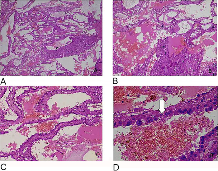
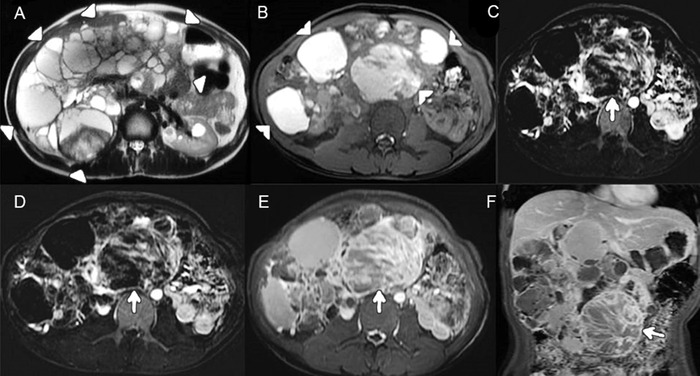
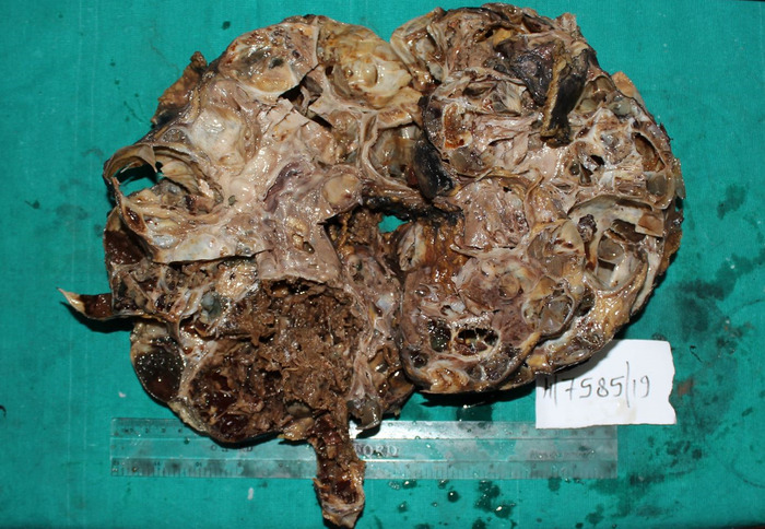
Background: Tubulocystic renal cell carcinoma is a lesser-known neoplastic entity compared to other common histologic variants of renal cell carcinoma. The World Health Organization identified tubulocystic renal cell carcinoma as a newly recognized renal tumor in 2016. We report a case of tubulocystic renal cell carcinoma in a young adult. Case Report: A 21-year-old male presented with the chief complaint of a lump on the right side of his abdomen since childhood. Magnetic resonance imaging revealed an enlarged right kidney with multiple large multiloculated cysts with hemorrhagic contents and enhancing peripheral nodular solid components with enhancing septa in some of the cysts, suggesting the possibility of multifocal intracystic papillary renal cell carcinoma. Imaging showed a Bosniak type IV cystic lesion in the right kidney. Right radical nephrectomy was performed. Grossly, the kidney was almost replaced by variable-sized cystic lesions with thick septations filled with serous fluid to gelatinous material. The tubules and cysts were lined by a single layer of flat, hobnail, and cuboidal cells with high-grade nuclear features. No ovarian-type stroma was identified. In places, a papillary component was also identified. Tubulocystic renal cell carcinoma was diagnosed based on microscopy and immunohistochemistry results. Conclusion: Tubulocystic renal cell carcinoma is a rare entity that was previously called Bellini duct carcinoma and low-grade collecting duct carcinoma. Because of the limited number of cases reported, tubulocystic renal cell carcinoma needs to be followed to determine the biologic behavior and metastatic potential of these tumors so that management guidelines for such cases can be developed.
期刊介绍:
The Ochsner Journal is a quarterly publication designed to support Ochsner"s mission to improve the health of our community through a commitment to innovation in healthcare, medical research, and education. The Ochsner Journal provides an active dialogue on practice standards in today"s changing healthcare environment. Emphasis will be given to topics of great societal and medical significance.

 求助内容:
求助内容: 应助结果提醒方式:


