Jia Liu, Lei Liu, Yang Su, Yi Wang, Yuchun Zhu, Xiaobin Sun, Yuanbiao Guo, Jing Shan
{"title":"IL-33参与食管腺癌的发生发展。","authors":"Jia Liu, Lei Liu, Yang Su, Yi Wang, Yuchun Zhu, Xiaobin Sun, Yuanbiao Guo, Jing Shan","doi":"10.3389/pore.2022.1610474","DOIUrl":null,"url":null,"abstract":"<p><p><b>Background:</b> The progression from chronic gastroesophageal reflux disease (GERD) to Barrett esophagus (BE) and esophageal adenocarcinoma (EAC) is an inflammatory-driven neoplastic change. Interleukin-33 (IL-33) has identified as a crucial factor in several inflammatory disorders and malignancies. <b>Methods:</b> The high-density tissue microarray of the human EAC was analyzed with IL-33 immunohistochemistry staining (IHC). By anastomosing the jejunum with the esophagus, the rat model of EAC with mixed gastroduodenal reflux was established. The expression of IL-33 was determined using quantitative real-time polymerase chain reaction (RT-qPCR), western blot (WB), IHC and enzyme-linked immunosorbent assay (ELISA). Esophageal adenocarcinoma cells (OE19 and OE33) and human esophageal epithelial cells (HEECs) were used. <b>Results:</b> In the cytoplasm of human EAC tissue, IL-33 expression was substantially greater than in adjacent normal tissue. In rat model, the expression of IL-33 in the EAC group was considerably greater than in the control group, and this expression increased with the upgrade of pathological stage. In <i>in vitro</i> experiment, the mRNA and protein levels of IL-33 were considerably greater in OE19 and OE33 than in HEECs. The stimulation of IL-33 enhanced the proliferation, migration, invasion, and epithelial-mesenchymal transition (EMT) of OE19 and OE33, but soluble ST2 (sST2) inhibited these effects. IL-33 stimulated the release of IL-6 by OE19 and OE33 cells. <b>Conclusion:</b> This study demonstrated the overexpression of IL-33 in the transition from GERD to EAC and that IL-33 promoted carcinogenesis in EAC cells through ST2. IL-33 might be a possible preventive target for EAC.</p>","PeriodicalId":411887,"journal":{"name":"Pathology oncology research : POR","volume":" ","pages":"1610474"},"PeriodicalIF":0.0000,"publicationDate":"2022-08-30","publicationTypes":"Journal Article","fieldsOfStudy":null,"isOpenAccess":false,"openAccessPdf":"https://www.ncbi.nlm.nih.gov/pmc/articles/PMC9469785/pdf/","citationCount":"2","resultStr":"{\"title\":\"IL-33 Participates in the Development of Esophageal Adenocarcinoma.\",\"authors\":\"Jia Liu, Lei Liu, Yang Su, Yi Wang, Yuchun Zhu, Xiaobin Sun, Yuanbiao Guo, Jing Shan\",\"doi\":\"10.3389/pore.2022.1610474\",\"DOIUrl\":null,\"url\":null,\"abstract\":\"<p><p><b>Background:</b> The progression from chronic gastroesophageal reflux disease (GERD) to Barrett esophagus (BE) and esophageal adenocarcinoma (EAC) is an inflammatory-driven neoplastic change. Interleukin-33 (IL-33) has identified as a crucial factor in several inflammatory disorders and malignancies. <b>Methods:</b> The high-density tissue microarray of the human EAC was analyzed with IL-33 immunohistochemistry staining (IHC). By anastomosing the jejunum with the esophagus, the rat model of EAC with mixed gastroduodenal reflux was established. The expression of IL-33 was determined using quantitative real-time polymerase chain reaction (RT-qPCR), western blot (WB), IHC and enzyme-linked immunosorbent assay (ELISA). Esophageal adenocarcinoma cells (OE19 and OE33) and human esophageal epithelial cells (HEECs) were used. <b>Results:</b> In the cytoplasm of human EAC tissue, IL-33 expression was substantially greater than in adjacent normal tissue. In rat model, the expression of IL-33 in the EAC group was considerably greater than in the control group, and this expression increased with the upgrade of pathological stage. In <i>in vitro</i> experiment, the mRNA and protein levels of IL-33 were considerably greater in OE19 and OE33 than in HEECs. The stimulation of IL-33 enhanced the proliferation, migration, invasion, and epithelial-mesenchymal transition (EMT) of OE19 and OE33, but soluble ST2 (sST2) inhibited these effects. IL-33 stimulated the release of IL-6 by OE19 and OE33 cells. <b>Conclusion:</b> This study demonstrated the overexpression of IL-33 in the transition from GERD to EAC and that IL-33 promoted carcinogenesis in EAC cells through ST2. IL-33 might be a possible preventive target for EAC.</p>\",\"PeriodicalId\":411887,\"journal\":{\"name\":\"Pathology oncology research : POR\",\"volume\":\" \",\"pages\":\"1610474\"},\"PeriodicalIF\":0.0000,\"publicationDate\":\"2022-08-30\",\"publicationTypes\":\"Journal Article\",\"fieldsOfStudy\":null,\"isOpenAccess\":false,\"openAccessPdf\":\"https://www.ncbi.nlm.nih.gov/pmc/articles/PMC9469785/pdf/\",\"citationCount\":\"2\",\"resultStr\":null,\"platform\":\"Semanticscholar\",\"paperid\":null,\"PeriodicalName\":\"Pathology oncology research : POR\",\"FirstCategoryId\":\"3\",\"ListUrlMain\":\"https://doi.org/10.3389/pore.2022.1610474\",\"RegionNum\":0,\"RegionCategory\":null,\"ArticlePicture\":[],\"TitleCN\":null,\"AbstractTextCN\":null,\"PMCID\":null,\"EPubDate\":\"2022/1/1 0:00:00\",\"PubModel\":\"eCollection\",\"JCR\":\"\",\"JCRName\":\"\",\"Score\":null,\"Total\":0}","platform":"Semanticscholar","paperid":null,"PeriodicalName":"Pathology oncology research : POR","FirstCategoryId":"3","ListUrlMain":"https://doi.org/10.3389/pore.2022.1610474","RegionNum":0,"RegionCategory":null,"ArticlePicture":[],"TitleCN":null,"AbstractTextCN":null,"PMCID":null,"EPubDate":"2022/1/1 0:00:00","PubModel":"eCollection","JCR":"","JCRName":"","Score":null,"Total":0}
引用次数: 2
摘要
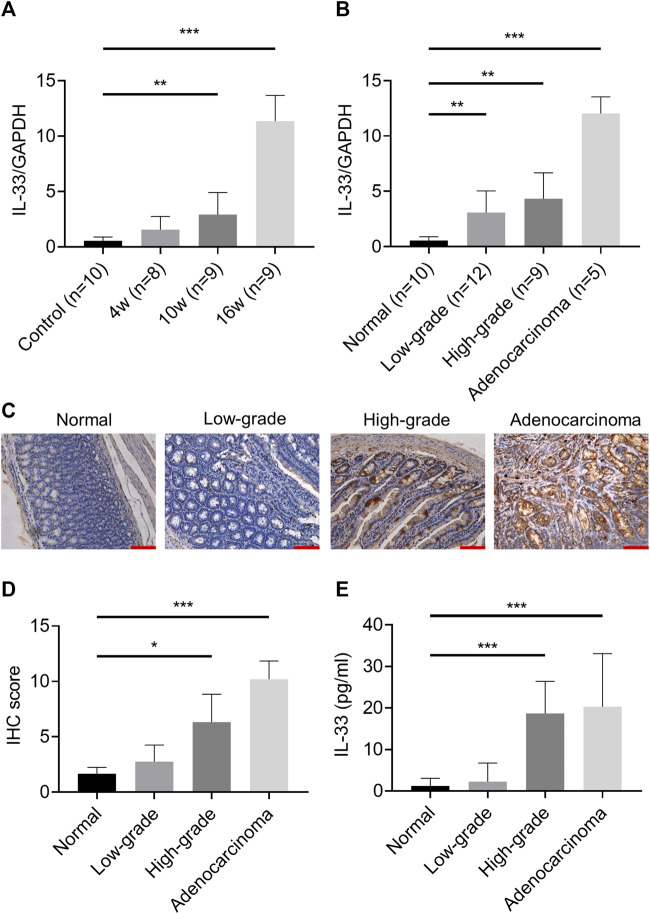
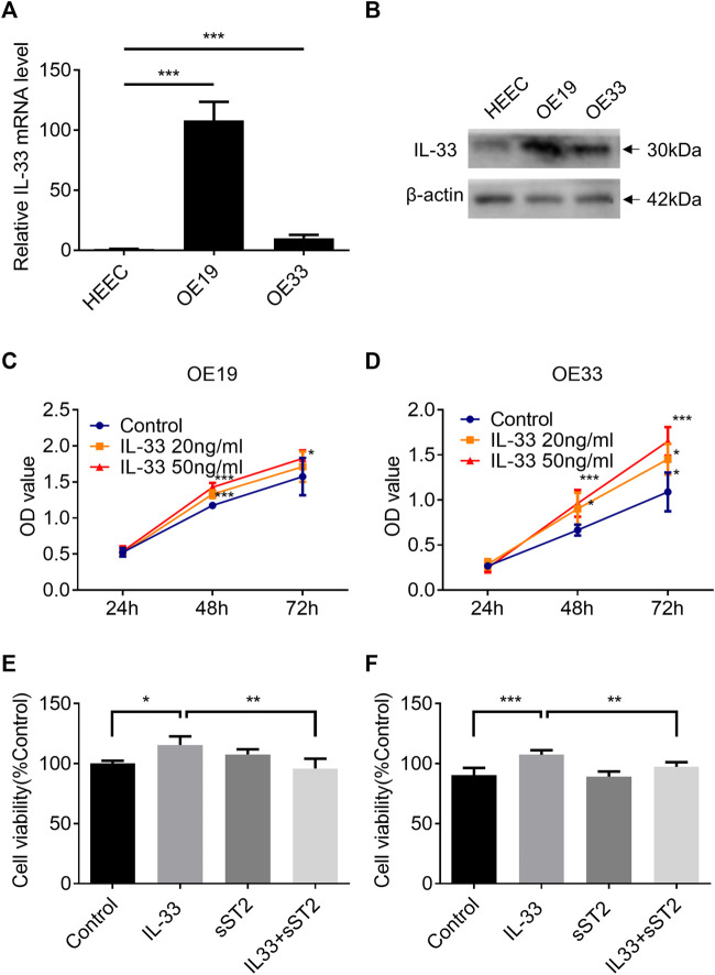
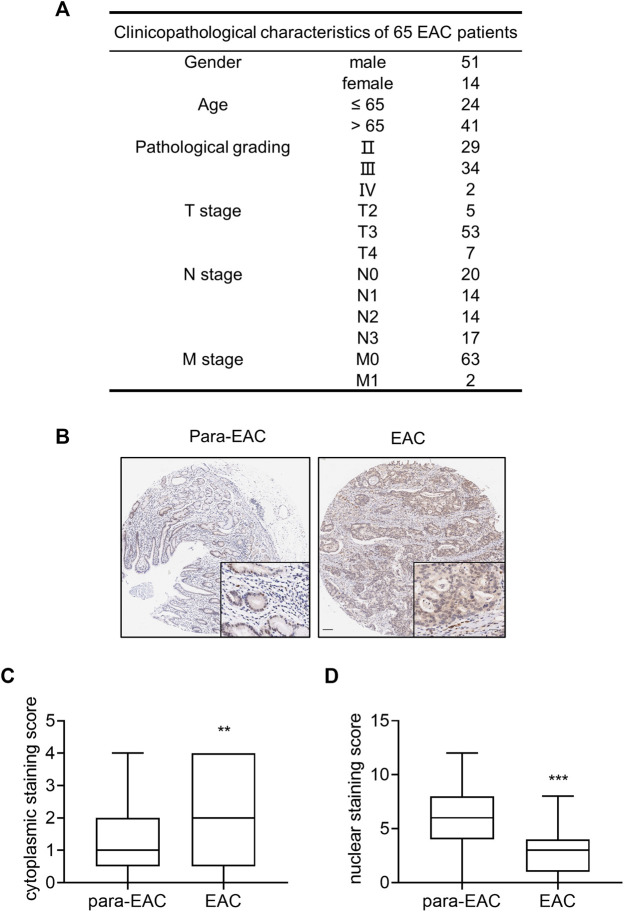
背景:从慢性胃食管反流病(GERD)发展到Barrett食管(BE)和食管腺癌(EAC)是一种炎症驱动的肿瘤改变。白细胞介素-33 (IL-33)已被确定为几种炎症性疾病和恶性肿瘤的关键因素。方法:采用IL-33免疫组化染色(IHC)对人EAC高密度组织芯片进行分析。通过将空肠与食管吻合,建立混合胃十二指肠反流大鼠EAC模型。采用实时荧光定量聚合酶链反应(RT-qPCR)、western blot (WB)、免疫组化(IHC)和酶联免疫吸附法(ELISA)检测IL-33的表达。采用食管腺癌细胞(OE19和OE33)和人食管上皮细胞(HEECs)。结果:人EAC组织细胞质中IL-33的表达明显高于邻近正常组织。在大鼠模型中,EAC组IL-33的表达明显高于对照组,且随着病理分期的升级而升高。在体外实验中,OE19和OE33中IL-33 mRNA和蛋白水平明显高于HEECs。IL-33的刺激增强了OE19和OE33的增殖、迁移、侵袭和上皮间质转化(EMT),但可溶性ST2 (sST2)抑制了这些作用。IL-33刺激OE19和OE33细胞释放IL-6。结论:本研究证实了IL-33在GERD向EAC过渡过程中过表达,IL-33通过ST2促进EAC细胞的癌变。IL-33可能是EAC的预防靶点。
IL-33 Participates in the Development of Esophageal Adenocarcinoma.
Background: The progression from chronic gastroesophageal reflux disease (GERD) to Barrett esophagus (BE) and esophageal adenocarcinoma (EAC) is an inflammatory-driven neoplastic change. Interleukin-33 (IL-33) has identified as a crucial factor in several inflammatory disorders and malignancies. Methods: The high-density tissue microarray of the human EAC was analyzed with IL-33 immunohistochemistry staining (IHC). By anastomosing the jejunum with the esophagus, the rat model of EAC with mixed gastroduodenal reflux was established. The expression of IL-33 was determined using quantitative real-time polymerase chain reaction (RT-qPCR), western blot (WB), IHC and enzyme-linked immunosorbent assay (ELISA). Esophageal adenocarcinoma cells (OE19 and OE33) and human esophageal epithelial cells (HEECs) were used. Results: In the cytoplasm of human EAC tissue, IL-33 expression was substantially greater than in adjacent normal tissue. In rat model, the expression of IL-33 in the EAC group was considerably greater than in the control group, and this expression increased with the upgrade of pathological stage. In in vitro experiment, the mRNA and protein levels of IL-33 were considerably greater in OE19 and OE33 than in HEECs. The stimulation of IL-33 enhanced the proliferation, migration, invasion, and epithelial-mesenchymal transition (EMT) of OE19 and OE33, but soluble ST2 (sST2) inhibited these effects. IL-33 stimulated the release of IL-6 by OE19 and OE33 cells. Conclusion: This study demonstrated the overexpression of IL-33 in the transition from GERD to EAC and that IL-33 promoted carcinogenesis in EAC cells through ST2. IL-33 might be a possible preventive target for EAC.

 求助内容:
求助内容: 应助结果提醒方式:


