Pablo Alejandro Rodriguez, Romina Chaintiou Piorno, Eugenia Pilar Consoli Lizzi
{"title":"牙髓显微外科治疗牙源性鼻窦病变。","authors":"Pablo Alejandro Rodriguez, Romina Chaintiou Piorno, Eugenia Pilar Consoli Lizzi","doi":"10.14740/jmc3982","DOIUrl":null,"url":null,"abstract":"<p><p>A case report is presented, in which a tooth with a chronic apical abscess that has caused a maxillary sinusitis was treated with an endodontic microsurgery. A 65-year-old female patient reported discomfort in a maxillary tooth gingiva. Clinically, a dental metal-ceramic bridge with adequate marginal adaptation, involving teeth from maxillary right canine to maxillary right second molar, was observed. The periapical radiograph showed the adequate adaptation of the crown and post in the maxillary right first premolar, an apical lesion, and the heavy thickening of the maxillary sinus floor mucosa. The diagnosis established in the maxillary right first premolar was of a previously treated tooth and a chronic apical abscess. Considering the accurate fixed prosthesis adaptation and the impossibility of disassembling the five-piece-metal-ceramic bridge to perform a new endodontic treatment and the prosthetic restoration, an endodontic microsurgery of this tooth was performed. The post-treatment follow-up of 12-month recall showed a normal thickness sinus membrane and a regenerating cortical bone. The proposed microsurgery has enabled the maintenance of the existing prosthetic rehabilitation of the tooth that caused the sinus pathosis.</p>","PeriodicalId":16279,"journal":{"name":"Journal of Medical Cases","volume":"13 9","pages":"456-461"},"PeriodicalIF":0.0000,"publicationDate":"2022-09-01","publicationTypes":"Journal Article","fieldsOfStudy":null,"isOpenAccess":false,"openAccessPdf":"https://ftp.ncbi.nlm.nih.gov/pub/pmc/oa_pdf/e7/9b/jmc-13-456.PMC9534196.pdf","citationCount":"1","resultStr":"{\"title\":\"Resolution of Odontogenic Sinus Pathosis by Endodontic Microsurgery.\",\"authors\":\"Pablo Alejandro Rodriguez, Romina Chaintiou Piorno, Eugenia Pilar Consoli Lizzi\",\"doi\":\"10.14740/jmc3982\",\"DOIUrl\":null,\"url\":null,\"abstract\":\"<p><p>A case report is presented, in which a tooth with a chronic apical abscess that has caused a maxillary sinusitis was treated with an endodontic microsurgery. A 65-year-old female patient reported discomfort in a maxillary tooth gingiva. Clinically, a dental metal-ceramic bridge with adequate marginal adaptation, involving teeth from maxillary right canine to maxillary right second molar, was observed. The periapical radiograph showed the adequate adaptation of the crown and post in the maxillary right first premolar, an apical lesion, and the heavy thickening of the maxillary sinus floor mucosa. The diagnosis established in the maxillary right first premolar was of a previously treated tooth and a chronic apical abscess. Considering the accurate fixed prosthesis adaptation and the impossibility of disassembling the five-piece-metal-ceramic bridge to perform a new endodontic treatment and the prosthetic restoration, an endodontic microsurgery of this tooth was performed. The post-treatment follow-up of 12-month recall showed a normal thickness sinus membrane and a regenerating cortical bone. The proposed microsurgery has enabled the maintenance of the existing prosthetic rehabilitation of the tooth that caused the sinus pathosis.</p>\",\"PeriodicalId\":16279,\"journal\":{\"name\":\"Journal of Medical Cases\",\"volume\":\"13 9\",\"pages\":\"456-461\"},\"PeriodicalIF\":0.0000,\"publicationDate\":\"2022-09-01\",\"publicationTypes\":\"Journal Article\",\"fieldsOfStudy\":null,\"isOpenAccess\":false,\"openAccessPdf\":\"https://ftp.ncbi.nlm.nih.gov/pub/pmc/oa_pdf/e7/9b/jmc-13-456.PMC9534196.pdf\",\"citationCount\":\"1\",\"resultStr\":null,\"platform\":\"Semanticscholar\",\"paperid\":null,\"PeriodicalName\":\"Journal of Medical Cases\",\"FirstCategoryId\":\"1085\",\"ListUrlMain\":\"https://doi.org/10.14740/jmc3982\",\"RegionNum\":0,\"RegionCategory\":null,\"ArticlePicture\":[],\"TitleCN\":null,\"AbstractTextCN\":null,\"PMCID\":null,\"EPubDate\":\"2022/9/28 0:00:00\",\"PubModel\":\"Epub\",\"JCR\":\"\",\"JCRName\":\"\",\"Score\":null,\"Total\":0}","platform":"Semanticscholar","paperid":null,"PeriodicalName":"Journal of Medical Cases","FirstCategoryId":"1085","ListUrlMain":"https://doi.org/10.14740/jmc3982","RegionNum":0,"RegionCategory":null,"ArticlePicture":[],"TitleCN":null,"AbstractTextCN":null,"PMCID":null,"EPubDate":"2022/9/28 0:00:00","PubModel":"Epub","JCR":"","JCRName":"","Score":null,"Total":0}
Resolution of Odontogenic Sinus Pathosis by Endodontic Microsurgery.
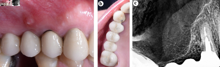
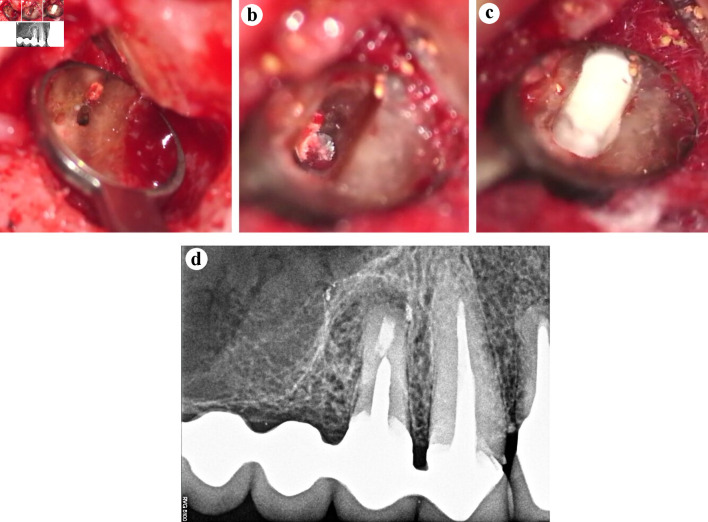
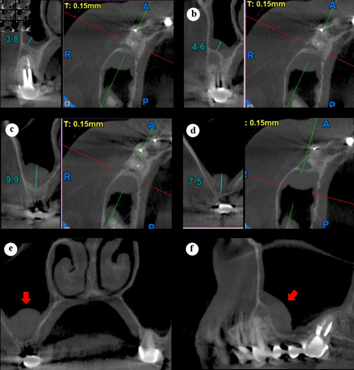
A case report is presented, in which a tooth with a chronic apical abscess that has caused a maxillary sinusitis was treated with an endodontic microsurgery. A 65-year-old female patient reported discomfort in a maxillary tooth gingiva. Clinically, a dental metal-ceramic bridge with adequate marginal adaptation, involving teeth from maxillary right canine to maxillary right second molar, was observed. The periapical radiograph showed the adequate adaptation of the crown and post in the maxillary right first premolar, an apical lesion, and the heavy thickening of the maxillary sinus floor mucosa. The diagnosis established in the maxillary right first premolar was of a previously treated tooth and a chronic apical abscess. Considering the accurate fixed prosthesis adaptation and the impossibility of disassembling the five-piece-metal-ceramic bridge to perform a new endodontic treatment and the prosthetic restoration, an endodontic microsurgery of this tooth was performed. The post-treatment follow-up of 12-month recall showed a normal thickness sinus membrane and a regenerating cortical bone. The proposed microsurgery has enabled the maintenance of the existing prosthetic rehabilitation of the tooth that caused the sinus pathosis.

 求助内容:
求助内容: 应助结果提醒方式:


