{"title":"扁桃体炎症性肌成纤维细胞瘤的病因分析和计算机断层成像:一个免疫能力强的病人的报告和简要回顾。","authors":"Yun-Zhen Luo, Li-Bo Dai, Shui-Hong Zhou, Xing-Mei Luo, Jun Fan, Ling-Xiang Ruan","doi":"10.1186/1758-3284-4-4","DOIUrl":null,"url":null,"abstract":"<p><strong>Objectives: </strong>The etiology of Inflammatory myofibroblastic tumor(IMT) is contentious. In this study, we used computed tomography (CT) to examine tonsillar IMT and further analyzed the etiology of this entity.</p><p><strong>Methodology: </strong>We presented CT features of left tonsillar IMT and reviewed the English-language literature published between 1984 and 2011.</p><p><strong>Results: </strong>To our knowledge, there are only six published cases of tonsillar IMT including the present case. Two patients were asymptomatic at initial presentation. Two patients were taking immunosuppressants, and one was pregnant and in an immunomodulated state. CT of our patient revealed a 2.6 × 1.8 cm irregular soft tissue mass between the left tonsil and the base of the tongue. It did not invade surrounding structures and was not enhanced on contrast-enhanced imaging.</p><p><strong>Conclusions: </strong>Tonsillar IMT may be a benign tumor. We suggest that preoperative recognition of tonsillar IMT by CT may be important to avoid unnecessary expanded surgery.</p>","PeriodicalId":49195,"journal":{"name":"Head and Neck Optical Diagnostics Society","volume":" ","pages":"4"},"PeriodicalIF":0.0000,"publicationDate":"2012-03-09","publicationTypes":"Journal Article","fieldsOfStudy":null,"isOpenAccess":false,"openAccessPdf":"https://sci-hub-pdf.com/10.1186/1758-3284-4-4","citationCount":"3","resultStr":"{\"title\":\"Etiology analysis and computed tomography imaging of a tonsillar inflammatory myofibroblastic tumor: report of an immunocompetent patient and brief review.\",\"authors\":\"Yun-Zhen Luo, Li-Bo Dai, Shui-Hong Zhou, Xing-Mei Luo, Jun Fan, Ling-Xiang Ruan\",\"doi\":\"10.1186/1758-3284-4-4\",\"DOIUrl\":null,\"url\":null,\"abstract\":\"<p><strong>Objectives: </strong>The etiology of Inflammatory myofibroblastic tumor(IMT) is contentious. In this study, we used computed tomography (CT) to examine tonsillar IMT and further analyzed the etiology of this entity.</p><p><strong>Methodology: </strong>We presented CT features of left tonsillar IMT and reviewed the English-language literature published between 1984 and 2011.</p><p><strong>Results: </strong>To our knowledge, there are only six published cases of tonsillar IMT including the present case. Two patients were asymptomatic at initial presentation. Two patients were taking immunosuppressants, and one was pregnant and in an immunomodulated state. CT of our patient revealed a 2.6 × 1.8 cm irregular soft tissue mass between the left tonsil and the base of the tongue. It did not invade surrounding structures and was not enhanced on contrast-enhanced imaging.</p><p><strong>Conclusions: </strong>Tonsillar IMT may be a benign tumor. We suggest that preoperative recognition of tonsillar IMT by CT may be important to avoid unnecessary expanded surgery.</p>\",\"PeriodicalId\":49195,\"journal\":{\"name\":\"Head and Neck Optical Diagnostics Society\",\"volume\":\" \",\"pages\":\"4\"},\"PeriodicalIF\":0.0000,\"publicationDate\":\"2012-03-09\",\"publicationTypes\":\"Journal Article\",\"fieldsOfStudy\":null,\"isOpenAccess\":false,\"openAccessPdf\":\"https://sci-hub-pdf.com/10.1186/1758-3284-4-4\",\"citationCount\":\"3\",\"resultStr\":null,\"platform\":\"Semanticscholar\",\"paperid\":null,\"PeriodicalName\":\"Head and Neck Optical Diagnostics Society\",\"FirstCategoryId\":\"1085\",\"ListUrlMain\":\"https://doi.org/10.1186/1758-3284-4-4\",\"RegionNum\":0,\"RegionCategory\":null,\"ArticlePicture\":[],\"TitleCN\":null,\"AbstractTextCN\":null,\"PMCID\":null,\"EPubDate\":\"\",\"PubModel\":\"\",\"JCR\":\"\",\"JCRName\":\"\",\"Score\":null,\"Total\":0}","platform":"Semanticscholar","paperid":null,"PeriodicalName":"Head and Neck Optical Diagnostics Society","FirstCategoryId":"1085","ListUrlMain":"https://doi.org/10.1186/1758-3284-4-4","RegionNum":0,"RegionCategory":null,"ArticlePicture":[],"TitleCN":null,"AbstractTextCN":null,"PMCID":null,"EPubDate":"","PubModel":"","JCR":"","JCRName":"","Score":null,"Total":0}
Etiology analysis and computed tomography imaging of a tonsillar inflammatory myofibroblastic tumor: report of an immunocompetent patient and brief review.
Objectives: The etiology of Inflammatory myofibroblastic tumor(IMT) is contentious. In this study, we used computed tomography (CT) to examine tonsillar IMT and further analyzed the etiology of this entity.
Methodology: We presented CT features of left tonsillar IMT and reviewed the English-language literature published between 1984 and 2011.
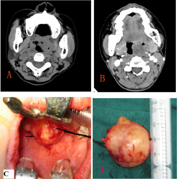
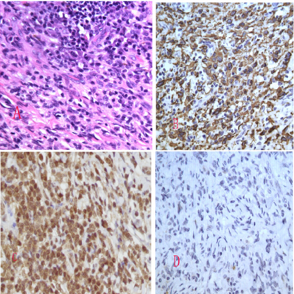
Results: To our knowledge, there are only six published cases of tonsillar IMT including the present case. Two patients were asymptomatic at initial presentation. Two patients were taking immunosuppressants, and one was pregnant and in an immunomodulated state. CT of our patient revealed a 2.6 × 1.8 cm irregular soft tissue mass between the left tonsil and the base of the tongue. It did not invade surrounding structures and was not enhanced on contrast-enhanced imaging.
Conclusions: Tonsillar IMT may be a benign tumor. We suggest that preoperative recognition of tonsillar IMT by CT may be important to avoid unnecessary expanded surgery.

 求助内容:
求助内容: 应助结果提醒方式:


