Jürgen Becker, Ana Covelo-Fernandez, Frederike von Bonin, Dieter Kube, Jörg Wilting
{"title":"鸡绒毛膜尿囊膜中ebv阳性伯基特淋巴瘤细胞特异性肿瘤-基质相互作用。","authors":"Jürgen Becker, Ana Covelo-Fernandez, Frederike von Bonin, Dieter Kube, Jörg Wilting","doi":"10.1186/2045-824X-4-3","DOIUrl":null,"url":null,"abstract":"<p><strong>Background: </strong>Burkitt's lymphoma (BL) is an aggressive Non-Hodgkin lymphoma. Epstein-Barr Virus (EBV) is able to transform B cells and is a causative infectious agent in BL. The precise role of EBV in lymphoma progression is still unclear. Most investigations have concentrated on cell autonomous functions of EBV in B cells. Functions of the local environment in BL progression have rarely been studied, mainly due to the lack of appropriate in vivo models. Therefore, we inoculated different human BL cell-lines onto the chorioallantoic membrane (CAM) of embryonic day 10 (ED10) chick embryos and re-incubated until ED14 and ED17.</p><p><strong>Results: </strong>All cell-lines formed solid tumors. However, we observed strong differences in the behavior of EBV+ and EBV- cell-lines. Tumor borders of EBV+ cells were very fuzzy and numerous cells migrated into the CAM. In EBV- tumors, the borders were much better defined. In contrast to EBV- cells, the EBV+ cells induced massive immigration of chick leukocytes at the tumor borders and the development of granulation tissue with large numbers of blood vessels and lymphatics, although the expression of pro- and anti-angiogenic forms of Vascular Endothelial Growth Factors/receptors was the same in all BL cell-lines tested. The EBV+ cell-lines massively disseminated via the lymphatics and completely occluded them.</p><p><strong>Conclusions: </strong>Our data suggest that the EBV+ cells attract pro-angiogenic leukocytes, which then induce secondary tumor-stroma interactions contributing to the progression of BL. We show that the CAM is a highly suitable in vivo model to study the differential behavior of BL cell-lines.</p>","PeriodicalId":23948,"journal":{"name":"Vascular Cell","volume":" ","pages":"3"},"PeriodicalIF":0.0000,"publicationDate":"2012-03-09","publicationTypes":"Journal Article","fieldsOfStudy":null,"isOpenAccess":false,"openAccessPdf":"https://sci-hub-pdf.com/10.1186/2045-824X-4-3","citationCount":"9","resultStr":"{\"title\":\"Specific tumor-stroma interactions of EBV-positive Burkitt's lymphoma cells in the chick chorioallantoic membrane.\",\"authors\":\"Jürgen Becker, Ana Covelo-Fernandez, Frederike von Bonin, Dieter Kube, Jörg Wilting\",\"doi\":\"10.1186/2045-824X-4-3\",\"DOIUrl\":null,\"url\":null,\"abstract\":\"<p><strong>Background: </strong>Burkitt's lymphoma (BL) is an aggressive Non-Hodgkin lymphoma. Epstein-Barr Virus (EBV) is able to transform B cells and is a causative infectious agent in BL. The precise role of EBV in lymphoma progression is still unclear. Most investigations have concentrated on cell autonomous functions of EBV in B cells. Functions of the local environment in BL progression have rarely been studied, mainly due to the lack of appropriate in vivo models. Therefore, we inoculated different human BL cell-lines onto the chorioallantoic membrane (CAM) of embryonic day 10 (ED10) chick embryos and re-incubated until ED14 and ED17.</p><p><strong>Results: </strong>All cell-lines formed solid tumors. However, we observed strong differences in the behavior of EBV+ and EBV- cell-lines. Tumor borders of EBV+ cells were very fuzzy and numerous cells migrated into the CAM. In EBV- tumors, the borders were much better defined. In contrast to EBV- cells, the EBV+ cells induced massive immigration of chick leukocytes at the tumor borders and the development of granulation tissue with large numbers of blood vessels and lymphatics, although the expression of pro- and anti-angiogenic forms of Vascular Endothelial Growth Factors/receptors was the same in all BL cell-lines tested. The EBV+ cell-lines massively disseminated via the lymphatics and completely occluded them.</p><p><strong>Conclusions: </strong>Our data suggest that the EBV+ cells attract pro-angiogenic leukocytes, which then induce secondary tumor-stroma interactions contributing to the progression of BL. We show that the CAM is a highly suitable in vivo model to study the differential behavior of BL cell-lines.</p>\",\"PeriodicalId\":23948,\"journal\":{\"name\":\"Vascular Cell\",\"volume\":\" \",\"pages\":\"3\"},\"PeriodicalIF\":0.0000,\"publicationDate\":\"2012-03-09\",\"publicationTypes\":\"Journal Article\",\"fieldsOfStudy\":null,\"isOpenAccess\":false,\"openAccessPdf\":\"https://sci-hub-pdf.com/10.1186/2045-824X-4-3\",\"citationCount\":\"9\",\"resultStr\":null,\"platform\":\"Semanticscholar\",\"paperid\":null,\"PeriodicalName\":\"Vascular Cell\",\"FirstCategoryId\":\"1085\",\"ListUrlMain\":\"https://doi.org/10.1186/2045-824X-4-3\",\"RegionNum\":0,\"RegionCategory\":null,\"ArticlePicture\":[],\"TitleCN\":null,\"AbstractTextCN\":null,\"PMCID\":null,\"EPubDate\":\"\",\"PubModel\":\"\",\"JCR\":\"Q4\",\"JCRName\":\"Neuroscience\",\"Score\":null,\"Total\":0}","platform":"Semanticscholar","paperid":null,"PeriodicalName":"Vascular Cell","FirstCategoryId":"1085","ListUrlMain":"https://doi.org/10.1186/2045-824X-4-3","RegionNum":0,"RegionCategory":null,"ArticlePicture":[],"TitleCN":null,"AbstractTextCN":null,"PMCID":null,"EPubDate":"","PubModel":"","JCR":"Q4","JCRName":"Neuroscience","Score":null,"Total":0}
Specific tumor-stroma interactions of EBV-positive Burkitt's lymphoma cells in the chick chorioallantoic membrane.
Background: Burkitt's lymphoma (BL) is an aggressive Non-Hodgkin lymphoma. Epstein-Barr Virus (EBV) is able to transform B cells and is a causative infectious agent in BL. The precise role of EBV in lymphoma progression is still unclear. Most investigations have concentrated on cell autonomous functions of EBV in B cells. Functions of the local environment in BL progression have rarely been studied, mainly due to the lack of appropriate in vivo models. Therefore, we inoculated different human BL cell-lines onto the chorioallantoic membrane (CAM) of embryonic day 10 (ED10) chick embryos and re-incubated until ED14 and ED17.
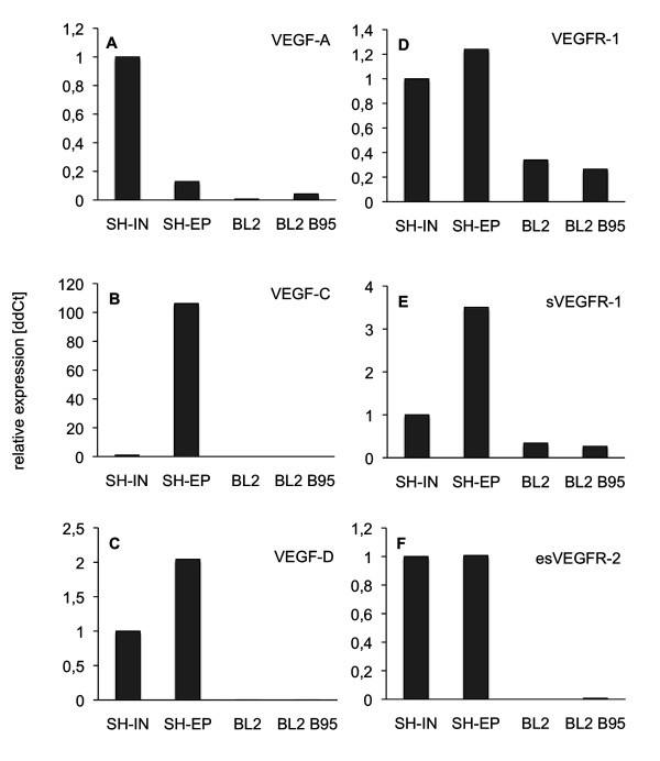
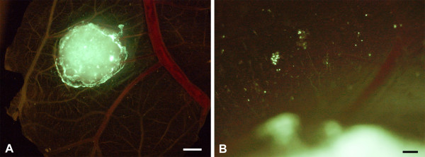
Results: All cell-lines formed solid tumors. However, we observed strong differences in the behavior of EBV+ and EBV- cell-lines. Tumor borders of EBV+ cells were very fuzzy and numerous cells migrated into the CAM. In EBV- tumors, the borders were much better defined. In contrast to EBV- cells, the EBV+ cells induced massive immigration of chick leukocytes at the tumor borders and the development of granulation tissue with large numbers of blood vessels and lymphatics, although the expression of pro- and anti-angiogenic forms of Vascular Endothelial Growth Factors/receptors was the same in all BL cell-lines tested. The EBV+ cell-lines massively disseminated via the lymphatics and completely occluded them.
Conclusions: Our data suggest that the EBV+ cells attract pro-angiogenic leukocytes, which then induce secondary tumor-stroma interactions contributing to the progression of BL. We show that the CAM is a highly suitable in vivo model to study the differential behavior of BL cell-lines.

 求助内容:
求助内容: 应助结果提醒方式:


