Fahmeed Hyder, Basavaraju G Sanganahalli, Peter Herman, Daniel Coman, Natasja J G Maandag, Kevin L Behar, Hal Blumenfeld, Douglas L Rothman
{"title":"动态校准功能磁共振成像中的神经血管和神经代谢耦合:块设计和事件相关范式的瞬时氧化神经能量学。","authors":"Fahmeed Hyder, Basavaraju G Sanganahalli, Peter Herman, Daniel Coman, Natasja J G Maandag, Kevin L Behar, Hal Blumenfeld, Douglas L Rothman","doi":"10.3389/fnene.2010.00018","DOIUrl":null,"url":null,"abstract":"<p><p>Functional magnetic resonance imaging (fMRI) with blood-oxygenation level dependent (BOLD) contrast is an important tool for mapping brain activity. Interest in quantitative fMRI has renewed awareness in importance of oxidative neuroenergetics, as reflected by cerebral metabolic rate of oxygen consumption(CMRO2), for supporting brain function. Relationships between BOLD signal and the underlying neurophysiological parameters have been elucidated to allow determination of dynamic changes inCMRO2 by \"calibrated fMRI,\" which require multi-modal measurements of BOLD signal along with cerebral blood flow (CBF) and volume (CBV). But how doCMRO2 changes, steady-state or transient, derived from calibrated fMRI compare with neural activity recordings of local field potential (LFP) and/or multi-unit activity (MUA)? Here we discuss recent findings primarily from animal studies which allow high magnetic fields studies for superior BOLD sensitivity as well as multi-modal CBV and CBF measurements in conjunction with LFP and MUA recordings from activated sites. A key observation is that while relationships between neural activity and sensory stimulus features range from linear to non-linear, associations between hyperemic components (BOLD, CBF, CBV) and neural activity (LFP, MUA) are almost always linear. More importantly, the results demonstrate good agreement between the changes inCMRO2 and independent measures of LFP or MUA. The tight neurovascular and neurometabolic couplings, observed from steady-state conditions to events separated by <200 ms, suggest rapid oxygen equilibration between blood and tissue pools and thus calibrated fMRI at high magnetic fields can provide high spatiotemporal mapping ofCMRO2 changes.</p>","PeriodicalId":88242,"journal":{"name":"Frontiers in neuroenergetics","volume":" ","pages":""},"PeriodicalIF":0.0000,"publicationDate":"2010-08-19","publicationTypes":"Journal Article","fieldsOfStudy":null,"isOpenAccess":false,"openAccessPdf":"https://sci-hub-pdf.com/10.3389/fnene.2010.00018","citationCount":"41","resultStr":"{\"title\":\"Neurovascular and Neurometabolic Couplings in Dynamic Calibrated fMRI: Transient Oxidative Neuroenergetics for Block-Design and Event-Related Paradigms.\",\"authors\":\"Fahmeed Hyder, Basavaraju G Sanganahalli, Peter Herman, Daniel Coman, Natasja J G Maandag, Kevin L Behar, Hal Blumenfeld, Douglas L Rothman\",\"doi\":\"10.3389/fnene.2010.00018\",\"DOIUrl\":null,\"url\":null,\"abstract\":\"<p><p>Functional magnetic resonance imaging (fMRI) with blood-oxygenation level dependent (BOLD) contrast is an important tool for mapping brain activity. Interest in quantitative fMRI has renewed awareness in importance of oxidative neuroenergetics, as reflected by cerebral metabolic rate of oxygen consumption(CMRO2), for supporting brain function. Relationships between BOLD signal and the underlying neurophysiological parameters have been elucidated to allow determination of dynamic changes inCMRO2 by \\\"calibrated fMRI,\\\" which require multi-modal measurements of BOLD signal along with cerebral blood flow (CBF) and volume (CBV). But how doCMRO2 changes, steady-state or transient, derived from calibrated fMRI compare with neural activity recordings of local field potential (LFP) and/or multi-unit activity (MUA)? Here we discuss recent findings primarily from animal studies which allow high magnetic fields studies for superior BOLD sensitivity as well as multi-modal CBV and CBF measurements in conjunction with LFP and MUA recordings from activated sites. A key observation is that while relationships between neural activity and sensory stimulus features range from linear to non-linear, associations between hyperemic components (BOLD, CBF, CBV) and neural activity (LFP, MUA) are almost always linear. More importantly, the results demonstrate good agreement between the changes inCMRO2 and independent measures of LFP or MUA. The tight neurovascular and neurometabolic couplings, observed from steady-state conditions to events separated by <200 ms, suggest rapid oxygen equilibration between blood and tissue pools and thus calibrated fMRI at high magnetic fields can provide high spatiotemporal mapping ofCMRO2 changes.</p>\",\"PeriodicalId\":88242,\"journal\":{\"name\":\"Frontiers in neuroenergetics\",\"volume\":\" \",\"pages\":\"\"},\"PeriodicalIF\":0.0000,\"publicationDate\":\"2010-08-19\",\"publicationTypes\":\"Journal Article\",\"fieldsOfStudy\":null,\"isOpenAccess\":false,\"openAccessPdf\":\"https://sci-hub-pdf.com/10.3389/fnene.2010.00018\",\"citationCount\":\"41\",\"resultStr\":null,\"platform\":\"Semanticscholar\",\"paperid\":null,\"PeriodicalName\":\"Frontiers in neuroenergetics\",\"FirstCategoryId\":\"1085\",\"ListUrlMain\":\"https://doi.org/10.3389/fnene.2010.00018\",\"RegionNum\":0,\"RegionCategory\":null,\"ArticlePicture\":[],\"TitleCN\":null,\"AbstractTextCN\":null,\"PMCID\":null,\"EPubDate\":\"2010/1/1 0:00:00\",\"PubModel\":\"eCollection\",\"JCR\":\"\",\"JCRName\":\"\",\"Score\":null,\"Total\":0}","platform":"Semanticscholar","paperid":null,"PeriodicalName":"Frontiers in neuroenergetics","FirstCategoryId":"1085","ListUrlMain":"https://doi.org/10.3389/fnene.2010.00018","RegionNum":0,"RegionCategory":null,"ArticlePicture":[],"TitleCN":null,"AbstractTextCN":null,"PMCID":null,"EPubDate":"2010/1/1 0:00:00","PubModel":"eCollection","JCR":"","JCRName":"","Score":null,"Total":0}
Neurovascular and Neurometabolic Couplings in Dynamic Calibrated fMRI: Transient Oxidative Neuroenergetics for Block-Design and Event-Related Paradigms.
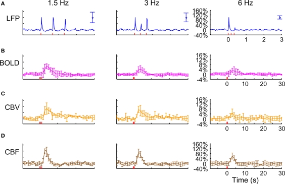
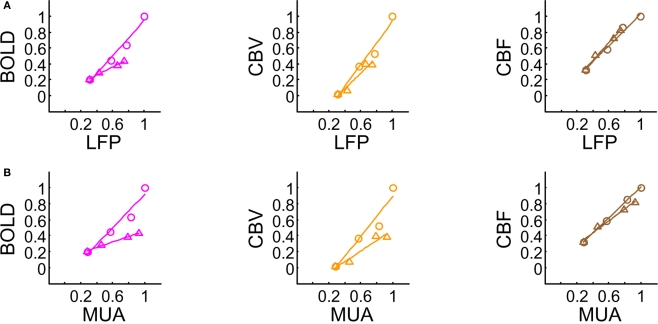
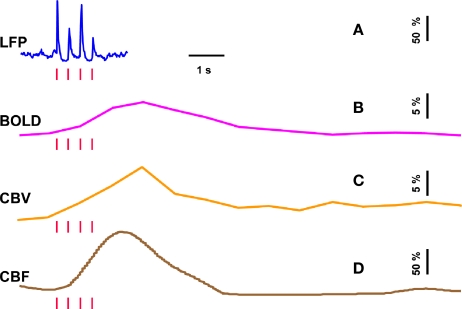
Functional magnetic resonance imaging (fMRI) with blood-oxygenation level dependent (BOLD) contrast is an important tool for mapping brain activity. Interest in quantitative fMRI has renewed awareness in importance of oxidative neuroenergetics, as reflected by cerebral metabolic rate of oxygen consumption(CMRO2), for supporting brain function. Relationships between BOLD signal and the underlying neurophysiological parameters have been elucidated to allow determination of dynamic changes inCMRO2 by "calibrated fMRI," which require multi-modal measurements of BOLD signal along with cerebral blood flow (CBF) and volume (CBV). But how doCMRO2 changes, steady-state or transient, derived from calibrated fMRI compare with neural activity recordings of local field potential (LFP) and/or multi-unit activity (MUA)? Here we discuss recent findings primarily from animal studies which allow high magnetic fields studies for superior BOLD sensitivity as well as multi-modal CBV and CBF measurements in conjunction with LFP and MUA recordings from activated sites. A key observation is that while relationships between neural activity and sensory stimulus features range from linear to non-linear, associations between hyperemic components (BOLD, CBF, CBV) and neural activity (LFP, MUA) are almost always linear. More importantly, the results demonstrate good agreement between the changes inCMRO2 and independent measures of LFP or MUA. The tight neurovascular and neurometabolic couplings, observed from steady-state conditions to events separated by <200 ms, suggest rapid oxygen equilibration between blood and tissue pools and thus calibrated fMRI at high magnetic fields can provide high spatiotemporal mapping ofCMRO2 changes.

 求助内容:
求助内容: 应助结果提醒方式:


