Francesco Latini, Markus Fahlström, David Fällmar, Niklas Marklund, Janet L Cunningham, Amalia Feresiadou
{"title":"弥散张量成像(DTI)在covid -19后自身免疫性脑炎中的表现是否优于标准磁共振成像(MRI) ?","authors":"Francesco Latini, Markus Fahlström, David Fällmar, Niklas Marklund, Janet L Cunningham, Amalia Feresiadou","doi":"10.48101/ujms.v127.8562","DOIUrl":null,"url":null,"abstract":"<p><strong>Background: </strong>Neurological and psychiatric manifestations related to severe acute respiratory syndrome coronavirus-2 (SARS-CoV-2) infection are widely recognised. Standard magnetic resonance imaging (MRI) investigations are normal in 40-80% of symptomatic patients, eventually delaying appropriate treatment when MRI is unrevealing any structural changes. The aim of this study is to investigate white matter abnormalities during an early stage of post-COVID-19 (coronavirus disease 2019) encephalitis while conventional MRI was normal.</p><p><strong>Methods: </strong>A patient with post-COVID-19 autoimmune encephalitis was investigated by serial MRIs and diffusion tensor imaging (DTI). Ten healthy control individuals (HC) were utilised as a control group for the DTI analysis. Major projection, commissural and association white matter pathways were reconstructed, and multiple diffusion parameters were analysed and then compared to the HC average using a z-test for serial examinations.</p><p><strong>Results: </strong>Eleven days after the onset of neurological symptoms, DTI revealed early white matter changes, compared with HC, when standard MRI was normal. On day 68, DTI showed multiple white matter lesions compared with HC, visible at this time also by the MRI images, indicating inflammatory changes in different association and projection white matter pathways.</p><p><strong>Conclusion: </strong>We confirm a limitation in the sensitivity of conventional MRI at the acute setting of post-COVID-19 autoimmune encephalitis. A complementary DTI investigation could be a valuable diagnostic tool in early therapeutic decisions concerning COVID-19-related neurological symptoms.</p>","PeriodicalId":23458,"journal":{"name":"Upsala journal of medical sciences","volume":" ","pages":""},"PeriodicalIF":1.6000,"publicationDate":"2022-05-10","publicationTypes":"Journal Article","fieldsOfStudy":null,"isOpenAccess":false,"openAccessPdf":"https://www.ncbi.nlm.nih.gov/pmc/articles/PMC9169543/pdf/","citationCount":"2","resultStr":"{\"title\":\"Can diffusion tensor imaging (DTI) outperform standard magnetic resonance imaging (MRI) investigations in post-COVID-19 autoimmune encephalitis?\",\"authors\":\"Francesco Latini, Markus Fahlström, David Fällmar, Niklas Marklund, Janet L Cunningham, Amalia Feresiadou\",\"doi\":\"10.48101/ujms.v127.8562\",\"DOIUrl\":null,\"url\":null,\"abstract\":\"<p><strong>Background: </strong>Neurological and psychiatric manifestations related to severe acute respiratory syndrome coronavirus-2 (SARS-CoV-2) infection are widely recognised. Standard magnetic resonance imaging (MRI) investigations are normal in 40-80% of symptomatic patients, eventually delaying appropriate treatment when MRI is unrevealing any structural changes. The aim of this study is to investigate white matter abnormalities during an early stage of post-COVID-19 (coronavirus disease 2019) encephalitis while conventional MRI was normal.</p><p><strong>Methods: </strong>A patient with post-COVID-19 autoimmune encephalitis was investigated by serial MRIs and diffusion tensor imaging (DTI). Ten healthy control individuals (HC) were utilised as a control group for the DTI analysis. Major projection, commissural and association white matter pathways were reconstructed, and multiple diffusion parameters were analysed and then compared to the HC average using a z-test for serial examinations.</p><p><strong>Results: </strong>Eleven days after the onset of neurological symptoms, DTI revealed early white matter changes, compared with HC, when standard MRI was normal. On day 68, DTI showed multiple white matter lesions compared with HC, visible at this time also by the MRI images, indicating inflammatory changes in different association and projection white matter pathways.</p><p><strong>Conclusion: </strong>We confirm a limitation in the sensitivity of conventional MRI at the acute setting of post-COVID-19 autoimmune encephalitis. A complementary DTI investigation could be a valuable diagnostic tool in early therapeutic decisions concerning COVID-19-related neurological symptoms.</p>\",\"PeriodicalId\":23458,\"journal\":{\"name\":\"Upsala journal of medical sciences\",\"volume\":\" \",\"pages\":\"\"},\"PeriodicalIF\":1.6000,\"publicationDate\":\"2022-05-10\",\"publicationTypes\":\"Journal Article\",\"fieldsOfStudy\":null,\"isOpenAccess\":false,\"openAccessPdf\":\"https://www.ncbi.nlm.nih.gov/pmc/articles/PMC9169543/pdf/\",\"citationCount\":\"2\",\"resultStr\":null,\"platform\":\"Semanticscholar\",\"paperid\":null,\"PeriodicalName\":\"Upsala journal of medical sciences\",\"FirstCategoryId\":\"3\",\"ListUrlMain\":\"https://doi.org/10.48101/ujms.v127.8562\",\"RegionNum\":4,\"RegionCategory\":\"医学\",\"ArticlePicture\":[],\"TitleCN\":null,\"AbstractTextCN\":null,\"PMCID\":null,\"EPubDate\":\"2022/1/1 0:00:00\",\"PubModel\":\"eCollection\",\"JCR\":\"Q2\",\"JCRName\":\"MEDICINE, GENERAL & INTERNAL\",\"Score\":null,\"Total\":0}","platform":"Semanticscholar","paperid":null,"PeriodicalName":"Upsala journal of medical sciences","FirstCategoryId":"3","ListUrlMain":"https://doi.org/10.48101/ujms.v127.8562","RegionNum":4,"RegionCategory":"医学","ArticlePicture":[],"TitleCN":null,"AbstractTextCN":null,"PMCID":null,"EPubDate":"2022/1/1 0:00:00","PubModel":"eCollection","JCR":"Q2","JCRName":"MEDICINE, GENERAL & INTERNAL","Score":null,"Total":0}
Can diffusion tensor imaging (DTI) outperform standard magnetic resonance imaging (MRI) investigations in post-COVID-19 autoimmune encephalitis?
Background: Neurological and psychiatric manifestations related to severe acute respiratory syndrome coronavirus-2 (SARS-CoV-2) infection are widely recognised. Standard magnetic resonance imaging (MRI) investigations are normal in 40-80% of symptomatic patients, eventually delaying appropriate treatment when MRI is unrevealing any structural changes. The aim of this study is to investigate white matter abnormalities during an early stage of post-COVID-19 (coronavirus disease 2019) encephalitis while conventional MRI was normal.
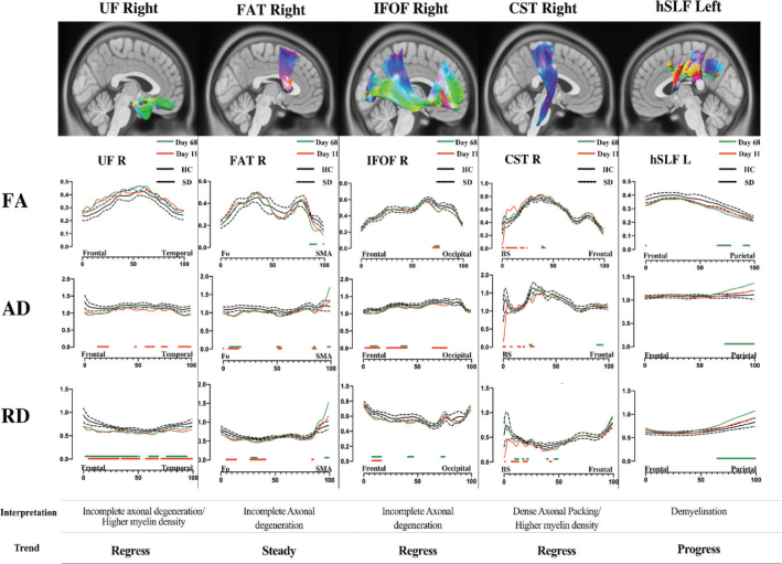
Methods: A patient with post-COVID-19 autoimmune encephalitis was investigated by serial MRIs and diffusion tensor imaging (DTI). Ten healthy control individuals (HC) were utilised as a control group for the DTI analysis. Major projection, commissural and association white matter pathways were reconstructed, and multiple diffusion parameters were analysed and then compared to the HC average using a z-test for serial examinations.
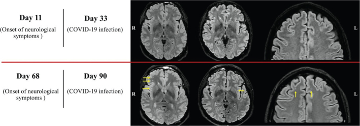
Results: Eleven days after the onset of neurological symptoms, DTI revealed early white matter changes, compared with HC, when standard MRI was normal. On day 68, DTI showed multiple white matter lesions compared with HC, visible at this time also by the MRI images, indicating inflammatory changes in different association and projection white matter pathways.
Conclusion: We confirm a limitation in the sensitivity of conventional MRI at the acute setting of post-COVID-19 autoimmune encephalitis. A complementary DTI investigation could be a valuable diagnostic tool in early therapeutic decisions concerning COVID-19-related neurological symptoms.
期刊介绍:
Upsala Journal of Medical Sciences is published for the Upsala Medical Society. It has been published since 1865 and is one of the oldest medical journals in Sweden.
The journal publishes clinical and experimental original works in the medical field. Although focusing on regional issues, the journal always welcomes contributions from outside Sweden.
Specially extended issues are published occasionally, dealing with special topics, congress proceedings and academic dissertations.
 求助内容:
求助内容: 应助结果提醒方式:


