Hanan F Hammouda, Mohammad M Farag, Mervat M F El Deftar, M Abdel-Gabbar, Basant M Mohamed
{"title":"ce掺杂生物活性玻璃/胶原/壳聚糖纳米复合支架对兔骨髓间充质干细胞源性成骨细胞形态和增殖的影响","authors":"Hanan F Hammouda, Mohammad M Farag, Mervat M F El Deftar, M Abdel-Gabbar, Basant M Mohamed","doi":"10.1186/s43141-022-00302-x","DOIUrl":null,"url":null,"abstract":"<p><strong>Background: </strong>Cerium-containing materials have wide applications in the biomedical field, because of the mimetic catalytic activities of cerium. The study aims to deeply estimate the biocompatibility of different scaffolds based on Ce-doped nanobioactive glass, collagen, and chitosan using the first passage of rabbit bone marrow mesenchymal stem cells (BM-MSCs) directed to osteogenic lineage by direct and indirect approach. One percentage of glass filler was used (30 wt. %) in the scaffold, while the percentage of CeO<sub>2</sub> in the glass was ranged from 0 to 10 mol. %. Cytotoxicity was evaluated by monitoring of cell morphological changes and reduction in cell proliferation activity of BMMSCs maintained under osteogenic condition using proliferation assays, MTT assay for the direct contact of cells/scaffolds twice in a week, trypan blue and hemocytometer cell counting for indirect contact of cells/scaffolds extracts at day 7. Cell behaviors growth, morphology characteristics were monitored daily under a microscope and cell counting were conducted after 1 week of the incubation of the cells with the extracts of the four composite scaffolds in the osteogenic medium at the end of the week.</p><p><strong>Results: </strong>Showed that at 24 h after direct contact with composite scaffold, all scaffolds showed proliferation of cells > 50% and increased in cell density on day 7. The scaffold of the highest percentage of CeO<sub>2</sub> in bioactive glass nanoparticles (sample CL/CH/C10) showed the lowest inhibition of cell proliferation (< 25%) at day 7. Moreover, the indirect cell viability test showed that all extracts from the four composite scaffolds did not demonstrate a toxic effect on the cells (inhibition value < 25%).</p><p><strong>Conclusion: </strong>The addition of CeO<sub>2</sub> to the glass composition improved the biocompatibility of the composite scaffold for the proliferation of rabbit bone marrow mesenchymal stem cells directed to osteogenic lineage.</p>","PeriodicalId":74026,"journal":{"name":"Journal, genetic engineering & biotechnology","volume":" ","pages":"33"},"PeriodicalIF":2.8000,"publicationDate":"2022-02-21","publicationTypes":"Journal Article","fieldsOfStudy":null,"isOpenAccess":false,"openAccessPdf":"https://www.ncbi.nlm.nih.gov/pmc/articles/PMC8864049/pdf/","citationCount":"12","resultStr":"{\"title\":\"Effect of Ce-doped bioactive glass/collagen/chitosan nanocomposite scaffolds on the cell morphology and proliferation of rabbit's bone marrow mesenchymal stem cells-derived osteogenic cells.\",\"authors\":\"Hanan F Hammouda, Mohammad M Farag, Mervat M F El Deftar, M Abdel-Gabbar, Basant M Mohamed\",\"doi\":\"10.1186/s43141-022-00302-x\",\"DOIUrl\":null,\"url\":null,\"abstract\":\"<p><strong>Background: </strong>Cerium-containing materials have wide applications in the biomedical field, because of the mimetic catalytic activities of cerium. The study aims to deeply estimate the biocompatibility of different scaffolds based on Ce-doped nanobioactive glass, collagen, and chitosan using the first passage of rabbit bone marrow mesenchymal stem cells (BM-MSCs) directed to osteogenic lineage by direct and indirect approach. One percentage of glass filler was used (30 wt. %) in the scaffold, while the percentage of CeO<sub>2</sub> in the glass was ranged from 0 to 10 mol. %. Cytotoxicity was evaluated by monitoring of cell morphological changes and reduction in cell proliferation activity of BMMSCs maintained under osteogenic condition using proliferation assays, MTT assay for the direct contact of cells/scaffolds twice in a week, trypan blue and hemocytometer cell counting for indirect contact of cells/scaffolds extracts at day 7. Cell behaviors growth, morphology characteristics were monitored daily under a microscope and cell counting were conducted after 1 week of the incubation of the cells with the extracts of the four composite scaffolds in the osteogenic medium at the end of the week.</p><p><strong>Results: </strong>Showed that at 24 h after direct contact with composite scaffold, all scaffolds showed proliferation of cells > 50% and increased in cell density on day 7. The scaffold of the highest percentage of CeO<sub>2</sub> in bioactive glass nanoparticles (sample CL/CH/C10) showed the lowest inhibition of cell proliferation (< 25%) at day 7. Moreover, the indirect cell viability test showed that all extracts from the four composite scaffolds did not demonstrate a toxic effect on the cells (inhibition value < 25%).</p><p><strong>Conclusion: </strong>The addition of CeO<sub>2</sub> to the glass composition improved the biocompatibility of the composite scaffold for the proliferation of rabbit bone marrow mesenchymal stem cells directed to osteogenic lineage.</p>\",\"PeriodicalId\":74026,\"journal\":{\"name\":\"Journal, genetic engineering & biotechnology\",\"volume\":\" \",\"pages\":\"33\"},\"PeriodicalIF\":2.8000,\"publicationDate\":\"2022-02-21\",\"publicationTypes\":\"Journal Article\",\"fieldsOfStudy\":null,\"isOpenAccess\":false,\"openAccessPdf\":\"https://www.ncbi.nlm.nih.gov/pmc/articles/PMC8864049/pdf/\",\"citationCount\":\"12\",\"resultStr\":null,\"platform\":\"Semanticscholar\",\"paperid\":null,\"PeriodicalName\":\"Journal, genetic engineering & biotechnology\",\"FirstCategoryId\":\"1085\",\"ListUrlMain\":\"https://doi.org/10.1186/s43141-022-00302-x\",\"RegionNum\":0,\"RegionCategory\":null,\"ArticlePicture\":[],\"TitleCN\":null,\"AbstractTextCN\":null,\"PMCID\":null,\"EPubDate\":\"\",\"PubModel\":\"\",\"JCR\":\"Q2\",\"JCRName\":\"BIOTECHNOLOGY & APPLIED MICROBIOLOGY\",\"Score\":null,\"Total\":0}","platform":"Semanticscholar","paperid":null,"PeriodicalName":"Journal, genetic engineering & biotechnology","FirstCategoryId":"1085","ListUrlMain":"https://doi.org/10.1186/s43141-022-00302-x","RegionNum":0,"RegionCategory":null,"ArticlePicture":[],"TitleCN":null,"AbstractTextCN":null,"PMCID":null,"EPubDate":"","PubModel":"","JCR":"Q2","JCRName":"BIOTECHNOLOGY & APPLIED MICROBIOLOGY","Score":null,"Total":0}
Effect of Ce-doped bioactive glass/collagen/chitosan nanocomposite scaffolds on the cell morphology and proliferation of rabbit's bone marrow mesenchymal stem cells-derived osteogenic cells.
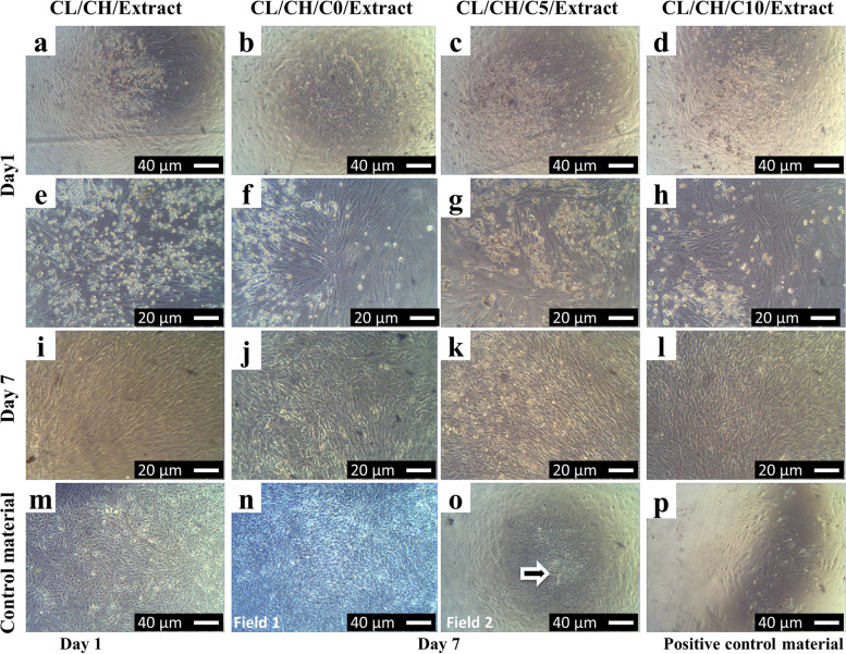
Background: Cerium-containing materials have wide applications in the biomedical field, because of the mimetic catalytic activities of cerium. The study aims to deeply estimate the biocompatibility of different scaffolds based on Ce-doped nanobioactive glass, collagen, and chitosan using the first passage of rabbit bone marrow mesenchymal stem cells (BM-MSCs) directed to osteogenic lineage by direct and indirect approach. One percentage of glass filler was used (30 wt. %) in the scaffold, while the percentage of CeO2 in the glass was ranged from 0 to 10 mol. %. Cytotoxicity was evaluated by monitoring of cell morphological changes and reduction in cell proliferation activity of BMMSCs maintained under osteogenic condition using proliferation assays, MTT assay for the direct contact of cells/scaffolds twice in a week, trypan blue and hemocytometer cell counting for indirect contact of cells/scaffolds extracts at day 7. Cell behaviors growth, morphology characteristics were monitored daily under a microscope and cell counting were conducted after 1 week of the incubation of the cells with the extracts of the four composite scaffolds in the osteogenic medium at the end of the week.
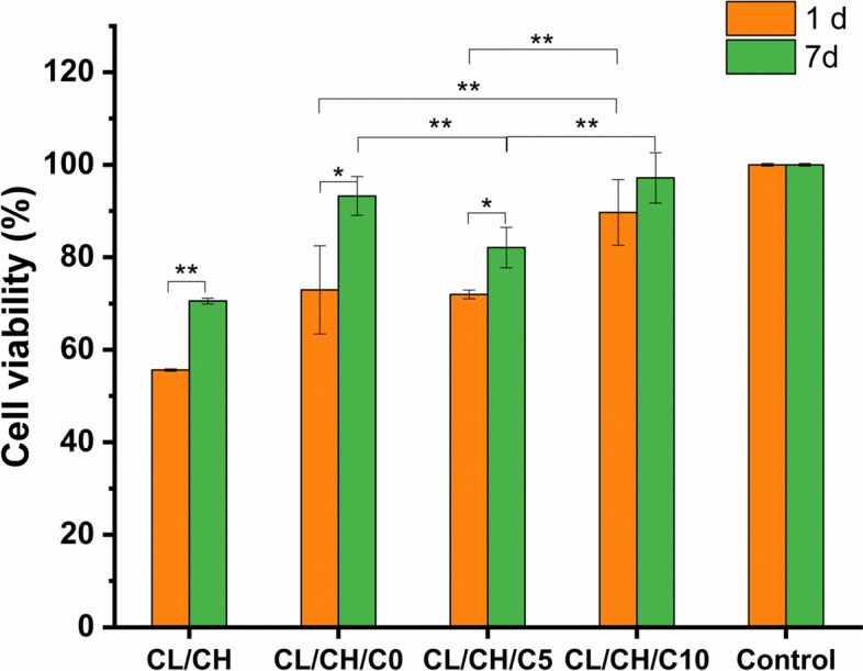
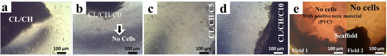
Results: Showed that at 24 h after direct contact with composite scaffold, all scaffolds showed proliferation of cells > 50% and increased in cell density on day 7. The scaffold of the highest percentage of CeO2 in bioactive glass nanoparticles (sample CL/CH/C10) showed the lowest inhibition of cell proliferation (< 25%) at day 7. Moreover, the indirect cell viability test showed that all extracts from the four composite scaffolds did not demonstrate a toxic effect on the cells (inhibition value < 25%).
Conclusion: The addition of CeO2 to the glass composition improved the biocompatibility of the composite scaffold for the proliferation of rabbit bone marrow mesenchymal stem cells directed to osteogenic lineage.

 求助内容:
求助内容: 应助结果提醒方式:


