Mira S Davidson, Clare Andradi-Brown, Sabrina Yahiya, Jill Chmielewski, Aidan J O'Donnell, Pratima Gurung, Myriam D Jeninga, Parichat Prommana, Dean W Andrew, Michaela Petter, Chairat Uthaipibull, Michelle J Boyle, George W Ashdown, Jeffrey D Dvorin, Sarah E Reece, Danny W Wilson, Kane A Cunningham, D Michael Ando, Michelle Dimon, Jake Baum
{"title":"利用卷积神经网络从细胞学涂片中自动检测和分期疟疾寄生虫。","authors":"Mira S Davidson, Clare Andradi-Brown, Sabrina Yahiya, Jill Chmielewski, Aidan J O'Donnell, Pratima Gurung, Myriam D Jeninga, Parichat Prommana, Dean W Andrew, Michaela Petter, Chairat Uthaipibull, Michelle J Boyle, George W Ashdown, Jeffrey D Dvorin, Sarah E Reece, Danny W Wilson, Kane A Cunningham, D Michael Ando, Michelle Dimon, Jake Baum","doi":"10.1017/S2633903X21000015","DOIUrl":null,"url":null,"abstract":"<p><p>Microscopic examination of blood smears remains the gold standard for laboratory inspection and diagnosis of malaria. Smear inspection is, however, time-consuming and dependent on trained microscopists with results varying in accuracy. We sought to develop an automated image analysis method to improve accuracy and standardization of smear inspection that retains capacity for expert confirmation and image archiving. Here, we present a machine learning method that achieves red blood cell (RBC) detection, differentiation between infected/uninfected cells, and parasite life stage categorization from unprocessed, heterogeneous smear images. Based on a pretrained Faster Region-Based Convolutional Neural Networks (R-CNN) model for RBC detection, our model performs accurately, with an average precision of 0.99 at an intersection-over-union threshold of 0.5. Application of a residual neural network-50 model to infected cells also performs accurately, with an area under the receiver operating characteristic curve of 0.98. Finally, combining our method with a regression model successfully recapitulates intraerythrocytic developmental cycle with accurate lifecycle stage categorization. Combined with a mobile-friendly web-based interface, called PlasmoCount, our method permits rapid navigation through and review of results for quality assurance. By standardizing assessment of Giemsa smears, our method markedly improves inspection reproducibility and presents a realistic route to both routine lab and future field-based automated malaria diagnosis.</p>","PeriodicalId":72371,"journal":{"name":"Biological imaging","volume":"1 ","pages":"e2"},"PeriodicalIF":0.0000,"publicationDate":"2021-08-02","publicationTypes":"Journal Article","fieldsOfStudy":null,"isOpenAccess":false,"openAccessPdf":"https://www.ncbi.nlm.nih.gov/pmc/articles/PMC8724263/pdf/","citationCount":"15","resultStr":"{\"title\":\"Automated detection and staging of malaria parasites from cytological smears using convolutional neural networks.\",\"authors\":\"Mira S Davidson, Clare Andradi-Brown, Sabrina Yahiya, Jill Chmielewski, Aidan J O'Donnell, Pratima Gurung, Myriam D Jeninga, Parichat Prommana, Dean W Andrew, Michaela Petter, Chairat Uthaipibull, Michelle J Boyle, George W Ashdown, Jeffrey D Dvorin, Sarah E Reece, Danny W Wilson, Kane A Cunningham, D Michael Ando, Michelle Dimon, Jake Baum\",\"doi\":\"10.1017/S2633903X21000015\",\"DOIUrl\":null,\"url\":null,\"abstract\":\"<p><p>Microscopic examination of blood smears remains the gold standard for laboratory inspection and diagnosis of malaria. Smear inspection is, however, time-consuming and dependent on trained microscopists with results varying in accuracy. We sought to develop an automated image analysis method to improve accuracy and standardization of smear inspection that retains capacity for expert confirmation and image archiving. Here, we present a machine learning method that achieves red blood cell (RBC) detection, differentiation between infected/uninfected cells, and parasite life stage categorization from unprocessed, heterogeneous smear images. Based on a pretrained Faster Region-Based Convolutional Neural Networks (R-CNN) model for RBC detection, our model performs accurately, with an average precision of 0.99 at an intersection-over-union threshold of 0.5. Application of a residual neural network-50 model to infected cells also performs accurately, with an area under the receiver operating characteristic curve of 0.98. Finally, combining our method with a regression model successfully recapitulates intraerythrocytic developmental cycle with accurate lifecycle stage categorization. Combined with a mobile-friendly web-based interface, called PlasmoCount, our method permits rapid navigation through and review of results for quality assurance. By standardizing assessment of Giemsa smears, our method markedly improves inspection reproducibility and presents a realistic route to both routine lab and future field-based automated malaria diagnosis.</p>\",\"PeriodicalId\":72371,\"journal\":{\"name\":\"Biological imaging\",\"volume\":\"1 \",\"pages\":\"e2\"},\"PeriodicalIF\":0.0000,\"publicationDate\":\"2021-08-02\",\"publicationTypes\":\"Journal Article\",\"fieldsOfStudy\":null,\"isOpenAccess\":false,\"openAccessPdf\":\"https://www.ncbi.nlm.nih.gov/pmc/articles/PMC8724263/pdf/\",\"citationCount\":\"15\",\"resultStr\":null,\"platform\":\"Semanticscholar\",\"paperid\":null,\"PeriodicalName\":\"Biological imaging\",\"FirstCategoryId\":\"1085\",\"ListUrlMain\":\"https://doi.org/10.1017/S2633903X21000015\",\"RegionNum\":0,\"RegionCategory\":null,\"ArticlePicture\":[],\"TitleCN\":null,\"AbstractTextCN\":null,\"PMCID\":null,\"EPubDate\":\"2021/1/1 0:00:00\",\"PubModel\":\"eCollection\",\"JCR\":\"\",\"JCRName\":\"\",\"Score\":null,\"Total\":0}","platform":"Semanticscholar","paperid":null,"PeriodicalName":"Biological imaging","FirstCategoryId":"1085","ListUrlMain":"https://doi.org/10.1017/S2633903X21000015","RegionNum":0,"RegionCategory":null,"ArticlePicture":[],"TitleCN":null,"AbstractTextCN":null,"PMCID":null,"EPubDate":"2021/1/1 0:00:00","PubModel":"eCollection","JCR":"","JCRName":"","Score":null,"Total":0}
Automated detection and staging of malaria parasites from cytological smears using convolutional neural networks.
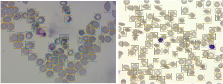
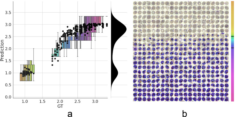
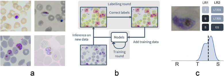
Microscopic examination of blood smears remains the gold standard for laboratory inspection and diagnosis of malaria. Smear inspection is, however, time-consuming and dependent on trained microscopists with results varying in accuracy. We sought to develop an automated image analysis method to improve accuracy and standardization of smear inspection that retains capacity for expert confirmation and image archiving. Here, we present a machine learning method that achieves red blood cell (RBC) detection, differentiation between infected/uninfected cells, and parasite life stage categorization from unprocessed, heterogeneous smear images. Based on a pretrained Faster Region-Based Convolutional Neural Networks (R-CNN) model for RBC detection, our model performs accurately, with an average precision of 0.99 at an intersection-over-union threshold of 0.5. Application of a residual neural network-50 model to infected cells also performs accurately, with an area under the receiver operating characteristic curve of 0.98. Finally, combining our method with a regression model successfully recapitulates intraerythrocytic developmental cycle with accurate lifecycle stage categorization. Combined with a mobile-friendly web-based interface, called PlasmoCount, our method permits rapid navigation through and review of results for quality assurance. By standardizing assessment of Giemsa smears, our method markedly improves inspection reproducibility and presents a realistic route to both routine lab and future field-based automated malaria diagnosis.

 求助内容:
求助内容: 应助结果提醒方式:


