Jeong-Yeon Kim, Jae-Hwan Choi, Eun Hye Oh, Seo-Young Choi, Hak-Jin Kim, Kwang-Dong Choi
{"title":"中枢神经系统淋巴瘤伴“双熊猫”征的孤立前庭综合征。","authors":"Jeong-Yeon Kim, Jae-Hwan Choi, Eun Hye Oh, Seo-Young Choi, Hak-Jin Kim, Kwang-Dong Choi","doi":"10.3988/jcn.2022.18.1.111","DOIUrl":null,"url":null,"abstract":"Dear Editor, Central nervous system (CNS) lymphoma is a rare type of non-Hodgkin lymphoma in which malignant cells from lymphoid tissue form in the brain and spinal cord (primary) or spread from other parts of the body to the brain and spinal cord (secondary).1 CNS lymphoma can manifest with various neurological symptoms depending on its location.2 A characteristic imaging finding of primary CNS lymphoma (PCNSL) is a computed tomography (CT) hyperdense enhanced supratentorial mass that is hypointense in T1-weighted magnetic resonance imaging (MRI) and isoto hypointense in T2-weighted MRI, with vivid homogeneous enhancement and restricted diffusion.1-3 We describe distinctive neuro-otological findings and the typical “double-panda” sign on brain MRI in a patient with CNS lymphoma. A 65-year-old male presented with acute dizziness with a 3-day history. Oculography showed horizontal gaze-evoked nystagmus (GEN) during bilateral gaze, and impaired horizontal smooth pursuit (Supplementary Fig. 1A in the online-only Data Supplement). Saccades were normal. Video head impulse tests (HITs) revealed decreased vestibulo-ocular reflex (VOR) gains of both horizontal and posterior semicircular canals (Supplementary Fig. 1B in the online-only Data Supplement). Ocular vestibular evoked myogenic potentials (VEMPs) were decreased during right-ear stimulation, while cervical VEMPs were symmetric (Supplementary Fig. 1C and D in the online-only Data Supplement). The levels of serum ceruloplasmin and 24-hour urine copper were normal, and Kayser-Fleischer rings were not observed. Brain MRI showed the characteristic “double-panda” sign with additional increased T2-weighted signal intensities along the bilateral ventricular walls, thalamus, hypothalamus, mammillary body, optic tract, dorsal pons, and upper medulla, and contrast enhancement in the ependyma (Fig. 1A-C). There was slight improvement of symptoms after treatment with steroid. One month later the patient was readmitted due to a sudden decrease in mentality. Brain CT showed obstructive hydrocephalus (Fig. 1F), and an external ventricular drain was placed. The endoscopic biopsy confirmed diffuse large-B-cell lymphoma (Supplementary Fig. 2 in the online-only Data Supplement). To the best of our knowledge, the present patient is unique since he showed the typical “double-panda” sign on brain MRI due to CNS lymphoma, which is traditionally considered to be characteristic of Wilson’s disease.4 The midbrain face of the “giant-panda” sign comprises normal-intensity red nuclei (eyes) and lateral portions of substantia nigra pars reticulata (ears), with high signal intensity in the tegmentum and hypointensity in the superior colliculus (mouth) (Fig. 1D). The pons face of the “miniature-panda” sign consists of relative hypointensity of the central tegmental tracts (eyes) contrasting with the hyperintensity of the aqueduct opening into the fourth ventricle (nose and mouth) (Fig. 1E).4 Combining these features produces the “double-panda” sign. Our case indicates that CNS lymphoma should be considered in the differential diagnosis of this rare imaging finding if there is no clear evidence of Wilson’s disease. CNS lymphoma in our patient was supported by the lack of evidence for toxic or metabolic Jeong-Yeon Kim* Jae-Hwan Choi* Eun Hye Oh Seo-Young Choi Hak-Jin Kim Kwang-Dong Choi","PeriodicalId":324902,"journal":{"name":"Journal of Clinical Neurology (Seoul, Korea)","volume":" ","pages":"111-113"},"PeriodicalIF":0.0000,"publicationDate":"2022-01-01","publicationTypes":"Journal Article","fieldsOfStudy":null,"isOpenAccess":false,"openAccessPdf":"https://ftp.ncbi.nlm.nih.gov/pub/pmc/oa_pdf/f4/69/jcn-18-111.PMC8762499.pdf","citationCount":"0","resultStr":"{\"title\":\"Isolated Vestibular Syndrome With \\\"Double-Panda\\\" Sign in CNS Lymphoma.\",\"authors\":\"Jeong-Yeon Kim, Jae-Hwan Choi, Eun Hye Oh, Seo-Young Choi, Hak-Jin Kim, Kwang-Dong Choi\",\"doi\":\"10.3988/jcn.2022.18.1.111\",\"DOIUrl\":null,\"url\":null,\"abstract\":\"Dear Editor, Central nervous system (CNS) lymphoma is a rare type of non-Hodgkin lymphoma in which malignant cells from lymphoid tissue form in the brain and spinal cord (primary) or spread from other parts of the body to the brain and spinal cord (secondary).1 CNS lymphoma can manifest with various neurological symptoms depending on its location.2 A characteristic imaging finding of primary CNS lymphoma (PCNSL) is a computed tomography (CT) hyperdense enhanced supratentorial mass that is hypointense in T1-weighted magnetic resonance imaging (MRI) and isoto hypointense in T2-weighted MRI, with vivid homogeneous enhancement and restricted diffusion.1-3 We describe distinctive neuro-otological findings and the typical “double-panda” sign on brain MRI in a patient with CNS lymphoma. A 65-year-old male presented with acute dizziness with a 3-day history. Oculography showed horizontal gaze-evoked nystagmus (GEN) during bilateral gaze, and impaired horizontal smooth pursuit (Supplementary Fig. 1A in the online-only Data Supplement). Saccades were normal. Video head impulse tests (HITs) revealed decreased vestibulo-ocular reflex (VOR) gains of both horizontal and posterior semicircular canals (Supplementary Fig. 1B in the online-only Data Supplement). Ocular vestibular evoked myogenic potentials (VEMPs) were decreased during right-ear stimulation, while cervical VEMPs were symmetric (Supplementary Fig. 1C and D in the online-only Data Supplement). The levels of serum ceruloplasmin and 24-hour urine copper were normal, and Kayser-Fleischer rings were not observed. Brain MRI showed the characteristic “double-panda” sign with additional increased T2-weighted signal intensities along the bilateral ventricular walls, thalamus, hypothalamus, mammillary body, optic tract, dorsal pons, and upper medulla, and contrast enhancement in the ependyma (Fig. 1A-C). There was slight improvement of symptoms after treatment with steroid. One month later the patient was readmitted due to a sudden decrease in mentality. Brain CT showed obstructive hydrocephalus (Fig. 1F), and an external ventricular drain was placed. The endoscopic biopsy confirmed diffuse large-B-cell lymphoma (Supplementary Fig. 2 in the online-only Data Supplement). To the best of our knowledge, the present patient is unique since he showed the typical “double-panda” sign on brain MRI due to CNS lymphoma, which is traditionally considered to be characteristic of Wilson’s disease.4 The midbrain face of the “giant-panda” sign comprises normal-intensity red nuclei (eyes) and lateral portions of substantia nigra pars reticulata (ears), with high signal intensity in the tegmentum and hypointensity in the superior colliculus (mouth) (Fig. 1D). The pons face of the “miniature-panda” sign consists of relative hypointensity of the central tegmental tracts (eyes) contrasting with the hyperintensity of the aqueduct opening into the fourth ventricle (nose and mouth) (Fig. 1E).4 Combining these features produces the “double-panda” sign. Our case indicates that CNS lymphoma should be considered in the differential diagnosis of this rare imaging finding if there is no clear evidence of Wilson’s disease. CNS lymphoma in our patient was supported by the lack of evidence for toxic or metabolic Jeong-Yeon Kim* Jae-Hwan Choi* Eun Hye Oh Seo-Young Choi Hak-Jin Kim Kwang-Dong Choi\",\"PeriodicalId\":324902,\"journal\":{\"name\":\"Journal of Clinical Neurology (Seoul, Korea)\",\"volume\":\" \",\"pages\":\"111-113\"},\"PeriodicalIF\":0.0000,\"publicationDate\":\"2022-01-01\",\"publicationTypes\":\"Journal Article\",\"fieldsOfStudy\":null,\"isOpenAccess\":false,\"openAccessPdf\":\"https://ftp.ncbi.nlm.nih.gov/pub/pmc/oa_pdf/f4/69/jcn-18-111.PMC8762499.pdf\",\"citationCount\":\"0\",\"resultStr\":null,\"platform\":\"Semanticscholar\",\"paperid\":null,\"PeriodicalName\":\"Journal of Clinical Neurology (Seoul, Korea)\",\"FirstCategoryId\":\"3\",\"ListUrlMain\":\"https://doi.org/10.3988/jcn.2022.18.1.111\",\"RegionNum\":0,\"RegionCategory\":null,\"ArticlePicture\":[],\"TitleCN\":null,\"AbstractTextCN\":null,\"PMCID\":null,\"EPubDate\":\"\",\"PubModel\":\"\",\"JCR\":\"\",\"JCRName\":\"\",\"Score\":null,\"Total\":0}","platform":"Semanticscholar","paperid":null,"PeriodicalName":"Journal of Clinical Neurology (Seoul, Korea)","FirstCategoryId":"3","ListUrlMain":"https://doi.org/10.3988/jcn.2022.18.1.111","RegionNum":0,"RegionCategory":null,"ArticlePicture":[],"TitleCN":null,"AbstractTextCN":null,"PMCID":null,"EPubDate":"","PubModel":"","JCR":"","JCRName":"","Score":null,"Total":0}
Isolated Vestibular Syndrome With "Double-Panda" Sign in CNS Lymphoma.
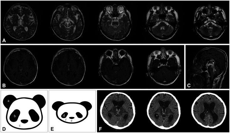
Dear Editor, Central nervous system (CNS) lymphoma is a rare type of non-Hodgkin lymphoma in which malignant cells from lymphoid tissue form in the brain and spinal cord (primary) or spread from other parts of the body to the brain and spinal cord (secondary).1 CNS lymphoma can manifest with various neurological symptoms depending on its location.2 A characteristic imaging finding of primary CNS lymphoma (PCNSL) is a computed tomography (CT) hyperdense enhanced supratentorial mass that is hypointense in T1-weighted magnetic resonance imaging (MRI) and isoto hypointense in T2-weighted MRI, with vivid homogeneous enhancement and restricted diffusion.1-3 We describe distinctive neuro-otological findings and the typical “double-panda” sign on brain MRI in a patient with CNS lymphoma. A 65-year-old male presented with acute dizziness with a 3-day history. Oculography showed horizontal gaze-evoked nystagmus (GEN) during bilateral gaze, and impaired horizontal smooth pursuit (Supplementary Fig. 1A in the online-only Data Supplement). Saccades were normal. Video head impulse tests (HITs) revealed decreased vestibulo-ocular reflex (VOR) gains of both horizontal and posterior semicircular canals (Supplementary Fig. 1B in the online-only Data Supplement). Ocular vestibular evoked myogenic potentials (VEMPs) were decreased during right-ear stimulation, while cervical VEMPs were symmetric (Supplementary Fig. 1C and D in the online-only Data Supplement). The levels of serum ceruloplasmin and 24-hour urine copper were normal, and Kayser-Fleischer rings were not observed. Brain MRI showed the characteristic “double-panda” sign with additional increased T2-weighted signal intensities along the bilateral ventricular walls, thalamus, hypothalamus, mammillary body, optic tract, dorsal pons, and upper medulla, and contrast enhancement in the ependyma (Fig. 1A-C). There was slight improvement of symptoms after treatment with steroid. One month later the patient was readmitted due to a sudden decrease in mentality. Brain CT showed obstructive hydrocephalus (Fig. 1F), and an external ventricular drain was placed. The endoscopic biopsy confirmed diffuse large-B-cell lymphoma (Supplementary Fig. 2 in the online-only Data Supplement). To the best of our knowledge, the present patient is unique since he showed the typical “double-panda” sign on brain MRI due to CNS lymphoma, which is traditionally considered to be characteristic of Wilson’s disease.4 The midbrain face of the “giant-panda” sign comprises normal-intensity red nuclei (eyes) and lateral portions of substantia nigra pars reticulata (ears), with high signal intensity in the tegmentum and hypointensity in the superior colliculus (mouth) (Fig. 1D). The pons face of the “miniature-panda” sign consists of relative hypointensity of the central tegmental tracts (eyes) contrasting with the hyperintensity of the aqueduct opening into the fourth ventricle (nose and mouth) (Fig. 1E).4 Combining these features produces the “double-panda” sign. Our case indicates that CNS lymphoma should be considered in the differential diagnosis of this rare imaging finding if there is no clear evidence of Wilson’s disease. CNS lymphoma in our patient was supported by the lack of evidence for toxic or metabolic Jeong-Yeon Kim* Jae-Hwan Choi* Eun Hye Oh Seo-Young Choi Hak-Jin Kim Kwang-Dong Choi

 求助内容:
求助内容: 应助结果提醒方式:


