Gopikrishna Deshpande, Yun Wang, Jennifer Robinson
{"title":"静息状态fMRI连接对人脑层流连接结构非常敏感。","authors":"Gopikrishna Deshpande, Yun Wang, Jennifer Robinson","doi":"10.1186/s40708-021-00150-4","DOIUrl":null,"url":null,"abstract":"<p><p>Previous invasive studies indicate that human neocortical graymatter contains cytoarchitectonically distinct layers, with notable differences in their structural connectivity with the rest of the brain. Given recent improvements in the spatial resolution of anatomical and functional magnetic resonance imaging (fMRI), we hypothesize that resting state functional connectivity (FC) derived from fMRI is sensitive to layer-specific thalamo-cortical and cortico-cortical microcircuits. Using sub-millimeter resting state fMRI data obtained at 7 T, we found that: (1) FC between the entire thalamus and cortical layers I and VI was significantly stronger than between the thalamus and other layers. Furthermore, FC between somatosensory thalamus (ventral posterolateral nucleus, VPL) and layers IV, VI of the primary somatosensory cortex were stronger than with other layers; (2) Inter-hemispheric cortico-cortical FC between homologous regions in superficial layers (layers I-III) was stronger compared to deep layers (layers V-VI). These findings are in agreement with structural connections inferred from previous invasive studies that showed that: (i) M-type neurons in the entire thalamus project to layer-I; (ii) Pyramidal neurons in layer-VI target all thalamic nuclei, (iii) C-type neurons in the VPL project to layer-IV and receive inputs from layer-VI of the primary somatosensory cortex, and (iv) 80% of collosal projecting neurons between homologous cortical regions connect superficial layers. Our results demonstrate for the first time that resting state fMRI is sensitive to structural connections between cortical layers (previously inferred through invasive studies), specifically in thalamo-cortical and cortico-cortical networks.</p>","PeriodicalId":37465,"journal":{"name":"Brain Informatics","volume":" ","pages":"2"},"PeriodicalIF":0.0000,"publicationDate":"2022-01-17","publicationTypes":"Journal Article","fieldsOfStudy":null,"isOpenAccess":false,"openAccessPdf":"https://www.ncbi.nlm.nih.gov/pmc/articles/PMC8764001/pdf/","citationCount":"3","resultStr":"{\"title\":\"Resting state fMRI connectivity is sensitive to laminar connectional architecture in the human brain.\",\"authors\":\"Gopikrishna Deshpande, Yun Wang, Jennifer Robinson\",\"doi\":\"10.1186/s40708-021-00150-4\",\"DOIUrl\":null,\"url\":null,\"abstract\":\"<p><p>Previous invasive studies indicate that human neocortical graymatter contains cytoarchitectonically distinct layers, with notable differences in their structural connectivity with the rest of the brain. Given recent improvements in the spatial resolution of anatomical and functional magnetic resonance imaging (fMRI), we hypothesize that resting state functional connectivity (FC) derived from fMRI is sensitive to layer-specific thalamo-cortical and cortico-cortical microcircuits. Using sub-millimeter resting state fMRI data obtained at 7 T, we found that: (1) FC between the entire thalamus and cortical layers I and VI was significantly stronger than between the thalamus and other layers. Furthermore, FC between somatosensory thalamus (ventral posterolateral nucleus, VPL) and layers IV, VI of the primary somatosensory cortex were stronger than with other layers; (2) Inter-hemispheric cortico-cortical FC between homologous regions in superficial layers (layers I-III) was stronger compared to deep layers (layers V-VI). These findings are in agreement with structural connections inferred from previous invasive studies that showed that: (i) M-type neurons in the entire thalamus project to layer-I; (ii) Pyramidal neurons in layer-VI target all thalamic nuclei, (iii) C-type neurons in the VPL project to layer-IV and receive inputs from layer-VI of the primary somatosensory cortex, and (iv) 80% of collosal projecting neurons between homologous cortical regions connect superficial layers. Our results demonstrate for the first time that resting state fMRI is sensitive to structural connections between cortical layers (previously inferred through invasive studies), specifically in thalamo-cortical and cortico-cortical networks.</p>\",\"PeriodicalId\":37465,\"journal\":{\"name\":\"Brain Informatics\",\"volume\":\" \",\"pages\":\"2\"},\"PeriodicalIF\":0.0000,\"publicationDate\":\"2022-01-17\",\"publicationTypes\":\"Journal Article\",\"fieldsOfStudy\":null,\"isOpenAccess\":false,\"openAccessPdf\":\"https://www.ncbi.nlm.nih.gov/pmc/articles/PMC8764001/pdf/\",\"citationCount\":\"3\",\"resultStr\":null,\"platform\":\"Semanticscholar\",\"paperid\":null,\"PeriodicalName\":\"Brain Informatics\",\"FirstCategoryId\":\"1085\",\"ListUrlMain\":\"https://doi.org/10.1186/s40708-021-00150-4\",\"RegionNum\":0,\"RegionCategory\":null,\"ArticlePicture\":[],\"TitleCN\":null,\"AbstractTextCN\":null,\"PMCID\":null,\"EPubDate\":\"\",\"PubModel\":\"\",\"JCR\":\"Q1\",\"JCRName\":\"Computer Science\",\"Score\":null,\"Total\":0}","platform":"Semanticscholar","paperid":null,"PeriodicalName":"Brain Informatics","FirstCategoryId":"1085","ListUrlMain":"https://doi.org/10.1186/s40708-021-00150-4","RegionNum":0,"RegionCategory":null,"ArticlePicture":[],"TitleCN":null,"AbstractTextCN":null,"PMCID":null,"EPubDate":"","PubModel":"","JCR":"Q1","JCRName":"Computer Science","Score":null,"Total":0}
Resting state fMRI connectivity is sensitive to laminar connectional architecture in the human brain.
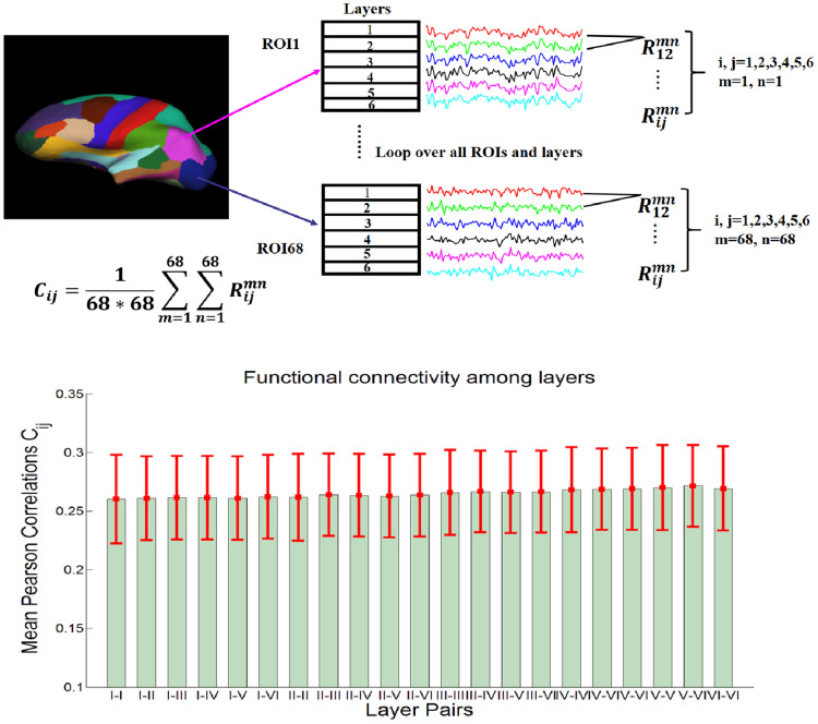
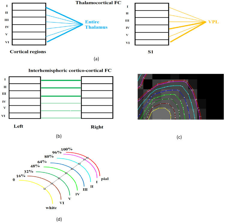
Previous invasive studies indicate that human neocortical graymatter contains cytoarchitectonically distinct layers, with notable differences in their structural connectivity with the rest of the brain. Given recent improvements in the spatial resolution of anatomical and functional magnetic resonance imaging (fMRI), we hypothesize that resting state functional connectivity (FC) derived from fMRI is sensitive to layer-specific thalamo-cortical and cortico-cortical microcircuits. Using sub-millimeter resting state fMRI data obtained at 7 T, we found that: (1) FC between the entire thalamus and cortical layers I and VI was significantly stronger than between the thalamus and other layers. Furthermore, FC between somatosensory thalamus (ventral posterolateral nucleus, VPL) and layers IV, VI of the primary somatosensory cortex were stronger than with other layers; (2) Inter-hemispheric cortico-cortical FC between homologous regions in superficial layers (layers I-III) was stronger compared to deep layers (layers V-VI). These findings are in agreement with structural connections inferred from previous invasive studies that showed that: (i) M-type neurons in the entire thalamus project to layer-I; (ii) Pyramidal neurons in layer-VI target all thalamic nuclei, (iii) C-type neurons in the VPL project to layer-IV and receive inputs from layer-VI of the primary somatosensory cortex, and (iv) 80% of collosal projecting neurons between homologous cortical regions connect superficial layers. Our results demonstrate for the first time that resting state fMRI is sensitive to structural connections between cortical layers (previously inferred through invasive studies), specifically in thalamo-cortical and cortico-cortical networks.
期刊介绍:
Brain Informatics is an international, peer-reviewed, interdisciplinary open-access journal published under the brand SpringerOpen, which provides a unique platform for researchers and practitioners to disseminate original research on computational and informatics technologies related to brain. This journal addresses the computational, cognitive, physiological, biological, physical, ecological and social perspectives of brain informatics. It also welcomes emerging information technologies and advanced neuro-imaging technologies, such as big data analytics and interactive knowledge discovery related to various large-scale brain studies and their applications. This journal will publish high-quality original research papers, brief reports and critical reviews in all theoretical, technological, clinical and interdisciplinary studies that make up the field of brain informatics and its applications in brain-machine intelligence, brain-inspired intelligent systems, mental health and brain disorders, etc. The scope of papers includes the following five tracks: Track 1: Cognitive and Computational Foundations of Brain Science Track 2: Human Information Processing Systems Track 3: Brain Big Data Analytics, Curation and Management Track 4: Informatics Paradigms for Brain and Mental Health Research Track 5: Brain-Machine Intelligence and Brain-Inspired Computing


 求助内容:
求助内容: 应助结果提醒方式:


