{"title":"EndMT:子宫内膜粘连纤维化中肌成纤维细胞起源的新发现。","authors":"Chengcheng Xu, Meng Bao, Xiaorong Fan, Jin Huang, Changhong Zhu, Wei Xia","doi":"10.1186/s12958-022-00887-5","DOIUrl":null,"url":null,"abstract":"<p><strong>Background: </strong>Intrauterine adhesion (IUA) is one of the leading causes of infertility and the main clinical challenge is the high recurrence rate. The key to solving this dilemma lies in elucidating the mechanisms of endometrial fibrosis. The aim of our team is to study the mechanism underlying intrauterine adhesion fibrosis and the origin of fibroblasts in the repair of endometrial fibrosis.</p><p><strong>Methods: </strong>Our experimental study involving an animal model of intrauterine adhesion and detection of fibrosis-related molecules. The levels of molecular factors related to the endothelial-to-mesenchymal transition (EndMT) were examined in a rat model of intrauterine adhesion using immunofluorescence, immunohistochemistry, qPCR and Western blot analyses. Main outcome measures are levels of the endothelial marker CD31 and the mesenchymal markers alpha-smooth muscle actin (α-SMA) and vimentin.</p><p><strong>Results: </strong>Immunofluorescence co-localization of CD31 and a-SMA showed that 14 days after moulding, double positive cells for CD31 and a-SMA could be clearly observed in the endometrium. Decreased CD31 levels and increased α-SMA and vimentin levels indicate that EndMT is involved in intrauterine adhesion fibrosis.</p><p><strong>Conclusions: </strong>Endothelial cells promote the emergence of fibroblasts via the EndMT during the endometrial fibrosis of intrauterine adhesions.</p>","PeriodicalId":520764,"journal":{"name":"Reproductive biology and endocrinology : RB&E","volume":" ","pages":"9"},"PeriodicalIF":0.0000,"publicationDate":"2022-01-07","publicationTypes":"Journal Article","fieldsOfStudy":null,"isOpenAccess":false,"openAccessPdf":"https://www.ncbi.nlm.nih.gov/pmc/articles/PMC8739974/pdf/","citationCount":"8","resultStr":"{\"title\":\"EndMT: New findings on the origin of myofibroblasts in endometrial fibrosis of intrauterine adhesions.\",\"authors\":\"Chengcheng Xu, Meng Bao, Xiaorong Fan, Jin Huang, Changhong Zhu, Wei Xia\",\"doi\":\"10.1186/s12958-022-00887-5\",\"DOIUrl\":null,\"url\":null,\"abstract\":\"<p><strong>Background: </strong>Intrauterine adhesion (IUA) is one of the leading causes of infertility and the main clinical challenge is the high recurrence rate. The key to solving this dilemma lies in elucidating the mechanisms of endometrial fibrosis. The aim of our team is to study the mechanism underlying intrauterine adhesion fibrosis and the origin of fibroblasts in the repair of endometrial fibrosis.</p><p><strong>Methods: </strong>Our experimental study involving an animal model of intrauterine adhesion and detection of fibrosis-related molecules. The levels of molecular factors related to the endothelial-to-mesenchymal transition (EndMT) were examined in a rat model of intrauterine adhesion using immunofluorescence, immunohistochemistry, qPCR and Western blot analyses. Main outcome measures are levels of the endothelial marker CD31 and the mesenchymal markers alpha-smooth muscle actin (α-SMA) and vimentin.</p><p><strong>Results: </strong>Immunofluorescence co-localization of CD31 and a-SMA showed that 14 days after moulding, double positive cells for CD31 and a-SMA could be clearly observed in the endometrium. Decreased CD31 levels and increased α-SMA and vimentin levels indicate that EndMT is involved in intrauterine adhesion fibrosis.</p><p><strong>Conclusions: </strong>Endothelial cells promote the emergence of fibroblasts via the EndMT during the endometrial fibrosis of intrauterine adhesions.</p>\",\"PeriodicalId\":520764,\"journal\":{\"name\":\"Reproductive biology and endocrinology : RB&E\",\"volume\":\" \",\"pages\":\"9\"},\"PeriodicalIF\":0.0000,\"publicationDate\":\"2022-01-07\",\"publicationTypes\":\"Journal Article\",\"fieldsOfStudy\":null,\"isOpenAccess\":false,\"openAccessPdf\":\"https://www.ncbi.nlm.nih.gov/pmc/articles/PMC8739974/pdf/\",\"citationCount\":\"8\",\"resultStr\":null,\"platform\":\"Semanticscholar\",\"paperid\":null,\"PeriodicalName\":\"Reproductive biology and endocrinology : RB&E\",\"FirstCategoryId\":\"3\",\"ListUrlMain\":\"https://doi.org/10.1186/s12958-022-00887-5\",\"RegionNum\":0,\"RegionCategory\":null,\"ArticlePicture\":[],\"TitleCN\":null,\"AbstractTextCN\":null,\"PMCID\":null,\"EPubDate\":\"\",\"PubModel\":\"\",\"JCR\":\"\",\"JCRName\":\"\",\"Score\":null,\"Total\":0}","platform":"Semanticscholar","paperid":null,"PeriodicalName":"Reproductive biology and endocrinology : RB&E","FirstCategoryId":"3","ListUrlMain":"https://doi.org/10.1186/s12958-022-00887-5","RegionNum":0,"RegionCategory":null,"ArticlePicture":[],"TitleCN":null,"AbstractTextCN":null,"PMCID":null,"EPubDate":"","PubModel":"","JCR":"","JCRName":"","Score":null,"Total":0}
EndMT: New findings on the origin of myofibroblasts in endometrial fibrosis of intrauterine adhesions.
Background: Intrauterine adhesion (IUA) is one of the leading causes of infertility and the main clinical challenge is the high recurrence rate. The key to solving this dilemma lies in elucidating the mechanisms of endometrial fibrosis. The aim of our team is to study the mechanism underlying intrauterine adhesion fibrosis and the origin of fibroblasts in the repair of endometrial fibrosis.
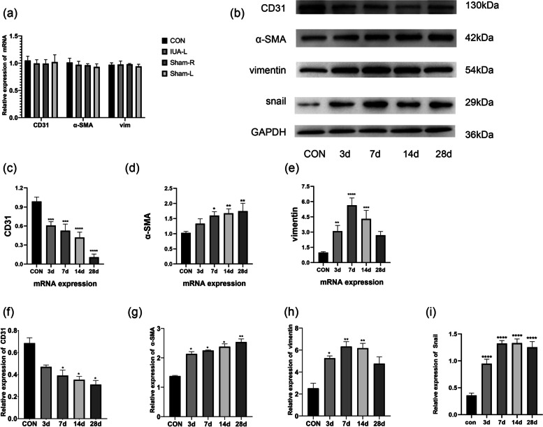
Methods: Our experimental study involving an animal model of intrauterine adhesion and detection of fibrosis-related molecules. The levels of molecular factors related to the endothelial-to-mesenchymal transition (EndMT) were examined in a rat model of intrauterine adhesion using immunofluorescence, immunohistochemistry, qPCR and Western blot analyses. Main outcome measures are levels of the endothelial marker CD31 and the mesenchymal markers alpha-smooth muscle actin (α-SMA) and vimentin.
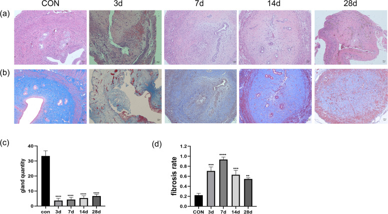
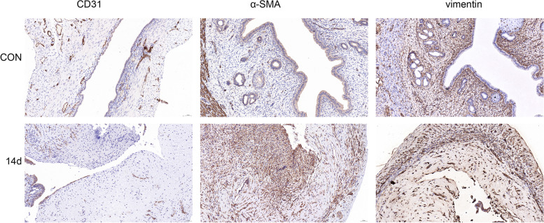
Results: Immunofluorescence co-localization of CD31 and a-SMA showed that 14 days after moulding, double positive cells for CD31 and a-SMA could be clearly observed in the endometrium. Decreased CD31 levels and increased α-SMA and vimentin levels indicate that EndMT is involved in intrauterine adhesion fibrosis.
Conclusions: Endothelial cells promote the emergence of fibroblasts via the EndMT during the endometrial fibrosis of intrauterine adhesions.

 求助内容:
求助内容: 应助结果提醒方式:


