{"title":"异常部位的总状脑室内神经囊虫病1例。","authors":"Priya Singh, Surya P Singh","doi":"10.4102/sajr.v25i1.2171","DOIUrl":null,"url":null,"abstract":"<p><p>Racemose and intraventricular neurocysticercosis are uncommon types of neurocysticercosis, resulting in a multiloculated, grape-like cluster appearance in the cerebrospinal fluid (CSF) spaces. A male patient presented with symptoms of raised intracranial pressure and demonstrated racemose neurocysticercosis at an atypical location involving the region of the crus of the fornix at the level of the body of lateral ventricles on magnetic resonance imaging. Associated intraventricular neurocysticercosis was seen in the atrium of the left lateral ventricle and fourth ventricle.</p>","PeriodicalId":43442,"journal":{"name":"SA Journal of Radiology","volume":null,"pages":null},"PeriodicalIF":0.7000,"publicationDate":"2021-12-06","publicationTypes":"Journal Article","fieldsOfStudy":null,"isOpenAccess":false,"openAccessPdf":"https://www.ncbi.nlm.nih.gov/pmc/articles/PMC8678948/pdf/","citationCount":"0","resultStr":"{\"title\":\"A case of racemose and intraventricular neurocysticercosis in an unusual location.\",\"authors\":\"Priya Singh, Surya P Singh\",\"doi\":\"10.4102/sajr.v25i1.2171\",\"DOIUrl\":null,\"url\":null,\"abstract\":\"<p><p>Racemose and intraventricular neurocysticercosis are uncommon types of neurocysticercosis, resulting in a multiloculated, grape-like cluster appearance in the cerebrospinal fluid (CSF) spaces. A male patient presented with symptoms of raised intracranial pressure and demonstrated racemose neurocysticercosis at an atypical location involving the region of the crus of the fornix at the level of the body of lateral ventricles on magnetic resonance imaging. Associated intraventricular neurocysticercosis was seen in the atrium of the left lateral ventricle and fourth ventricle.</p>\",\"PeriodicalId\":43442,\"journal\":{\"name\":\"SA Journal of Radiology\",\"volume\":null,\"pages\":null},\"PeriodicalIF\":0.7000,\"publicationDate\":\"2021-12-06\",\"publicationTypes\":\"Journal Article\",\"fieldsOfStudy\":null,\"isOpenAccess\":false,\"openAccessPdf\":\"https://www.ncbi.nlm.nih.gov/pmc/articles/PMC8678948/pdf/\",\"citationCount\":\"0\",\"resultStr\":null,\"platform\":\"Semanticscholar\",\"paperid\":null,\"PeriodicalName\":\"SA Journal of Radiology\",\"FirstCategoryId\":\"1085\",\"ListUrlMain\":\"https://doi.org/10.4102/sajr.v25i1.2171\",\"RegionNum\":0,\"RegionCategory\":null,\"ArticlePicture\":[],\"TitleCN\":null,\"AbstractTextCN\":null,\"PMCID\":null,\"EPubDate\":\"2021/1/1 0:00:00\",\"PubModel\":\"eCollection\",\"JCR\":\"Q4\",\"JCRName\":\"RADIOLOGY, NUCLEAR MEDICINE & MEDICAL IMAGING\",\"Score\":null,\"Total\":0}","platform":"Semanticscholar","paperid":null,"PeriodicalName":"SA Journal of Radiology","FirstCategoryId":"1085","ListUrlMain":"https://doi.org/10.4102/sajr.v25i1.2171","RegionNum":0,"RegionCategory":null,"ArticlePicture":[],"TitleCN":null,"AbstractTextCN":null,"PMCID":null,"EPubDate":"2021/1/1 0:00:00","PubModel":"eCollection","JCR":"Q4","JCRName":"RADIOLOGY, NUCLEAR MEDICINE & MEDICAL IMAGING","Score":null,"Total":0}
A case of racemose and intraventricular neurocysticercosis in an unusual location.
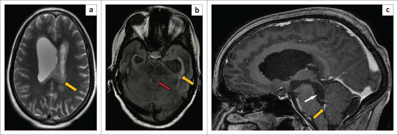
Racemose and intraventricular neurocysticercosis are uncommon types of neurocysticercosis, resulting in a multiloculated, grape-like cluster appearance in the cerebrospinal fluid (CSF) spaces. A male patient presented with symptoms of raised intracranial pressure and demonstrated racemose neurocysticercosis at an atypical location involving the region of the crus of the fornix at the level of the body of lateral ventricles on magnetic resonance imaging. Associated intraventricular neurocysticercosis was seen in the atrium of the left lateral ventricle and fourth ventricle.
期刊介绍:
The SA Journal of Radiology is the official journal of the Radiological Society of South Africa and the Professional Association of Radiologists in South Africa and Namibia. The SA Journal of Radiology is a general diagnostic radiological journal which carries original research and review articles, pictorial essays, case reports, letters, editorials, radiological practice and other radiological articles.