{"title":"分数各向异性揭示的人脑微观结构特性可以预测间歇性θ波脉冲刺激的后效。","authors":"Ikko Kimura, Hiroki Oishi, Masamichi J Hayashi, Kaoru Amano","doi":"10.1093/texcom/tgab065","DOIUrl":null,"url":null,"abstract":"<p><p>Intermittent theta burst stimulation (iTBS) delivered by transcranial magnetic stimulation (TMS) produces a long-term potentiation-like after-effect useful for investigations of cortical function and of potential therapeutic value. However, the iTBS after-effect over the primary motor cortex (M1) as measured by changes in motor evoked potential (MEP) amplitude exhibits a largely unexplained variability across individuals. Here, we present evidence that individual differences in white matter (WM) and gray matter (GM) microstructural properties revealed by fractional anisotropy (FA) predict the magnitude of the iTBS-induced after-effect over M1. The MEP amplitude change in the early phase (5-10 min post-iTBS) was associated with FA values in WM tracts such as right superior longitudinal fasciculus and corpus callosum. By contrast, the MEP amplitude change in the late phase (15-30 min post-iTBS) was associated with FA in GM, primarily in right frontal cortex. These results suggest that the microstructural properties of regions connected directly or indirectly to the target region (M1) are crucial determinants of the iTBS after-effect. FA values indicative of these microstructural differences can predict the potential effectiveness of repetitive TMS for both investigational use and clinical application.</p>","PeriodicalId":72551,"journal":{"name":"Cerebral cortex communications","volume":" ","pages":"tgab065"},"PeriodicalIF":0.0000,"publicationDate":"2021-12-15","publicationTypes":"Journal Article","fieldsOfStudy":null,"isOpenAccess":false,"openAccessPdf":"https://www.ncbi.nlm.nih.gov/pmc/articles/PMC8784864/pdf/","citationCount":"2","resultStr":"{\"title\":\"Microstructural Properties of Human Brain Revealed by Fractional Anisotropy Can Predict the After-Effect of Intermittent Theta Burst Stimulation.\",\"authors\":\"Ikko Kimura, Hiroki Oishi, Masamichi J Hayashi, Kaoru Amano\",\"doi\":\"10.1093/texcom/tgab065\",\"DOIUrl\":null,\"url\":null,\"abstract\":\"<p><p>Intermittent theta burst stimulation (iTBS) delivered by transcranial magnetic stimulation (TMS) produces a long-term potentiation-like after-effect useful for investigations of cortical function and of potential therapeutic value. However, the iTBS after-effect over the primary motor cortex (M1) as measured by changes in motor evoked potential (MEP) amplitude exhibits a largely unexplained variability across individuals. Here, we present evidence that individual differences in white matter (WM) and gray matter (GM) microstructural properties revealed by fractional anisotropy (FA) predict the magnitude of the iTBS-induced after-effect over M1. The MEP amplitude change in the early phase (5-10 min post-iTBS) was associated with FA values in WM tracts such as right superior longitudinal fasciculus and corpus callosum. By contrast, the MEP amplitude change in the late phase (15-30 min post-iTBS) was associated with FA in GM, primarily in right frontal cortex. These results suggest that the microstructural properties of regions connected directly or indirectly to the target region (M1) are crucial determinants of the iTBS after-effect. FA values indicative of these microstructural differences can predict the potential effectiveness of repetitive TMS for both investigational use and clinical application.</p>\",\"PeriodicalId\":72551,\"journal\":{\"name\":\"Cerebral cortex communications\",\"volume\":\" \",\"pages\":\"tgab065\"},\"PeriodicalIF\":0.0000,\"publicationDate\":\"2021-12-15\",\"publicationTypes\":\"Journal Article\",\"fieldsOfStudy\":null,\"isOpenAccess\":false,\"openAccessPdf\":\"https://www.ncbi.nlm.nih.gov/pmc/articles/PMC8784864/pdf/\",\"citationCount\":\"2\",\"resultStr\":null,\"platform\":\"Semanticscholar\",\"paperid\":null,\"PeriodicalName\":\"Cerebral cortex communications\",\"FirstCategoryId\":\"1085\",\"ListUrlMain\":\"https://doi.org/10.1093/texcom/tgab065\",\"RegionNum\":0,\"RegionCategory\":null,\"ArticlePicture\":[],\"TitleCN\":null,\"AbstractTextCN\":null,\"PMCID\":null,\"EPubDate\":\"2022/1/1 0:00:00\",\"PubModel\":\"eCollection\",\"JCR\":\"\",\"JCRName\":\"\",\"Score\":null,\"Total\":0}","platform":"Semanticscholar","paperid":null,"PeriodicalName":"Cerebral cortex communications","FirstCategoryId":"1085","ListUrlMain":"https://doi.org/10.1093/texcom/tgab065","RegionNum":0,"RegionCategory":null,"ArticlePicture":[],"TitleCN":null,"AbstractTextCN":null,"PMCID":null,"EPubDate":"2022/1/1 0:00:00","PubModel":"eCollection","JCR":"","JCRName":"","Score":null,"Total":0}
Microstructural Properties of Human Brain Revealed by Fractional Anisotropy Can Predict the After-Effect of Intermittent Theta Burst Stimulation.
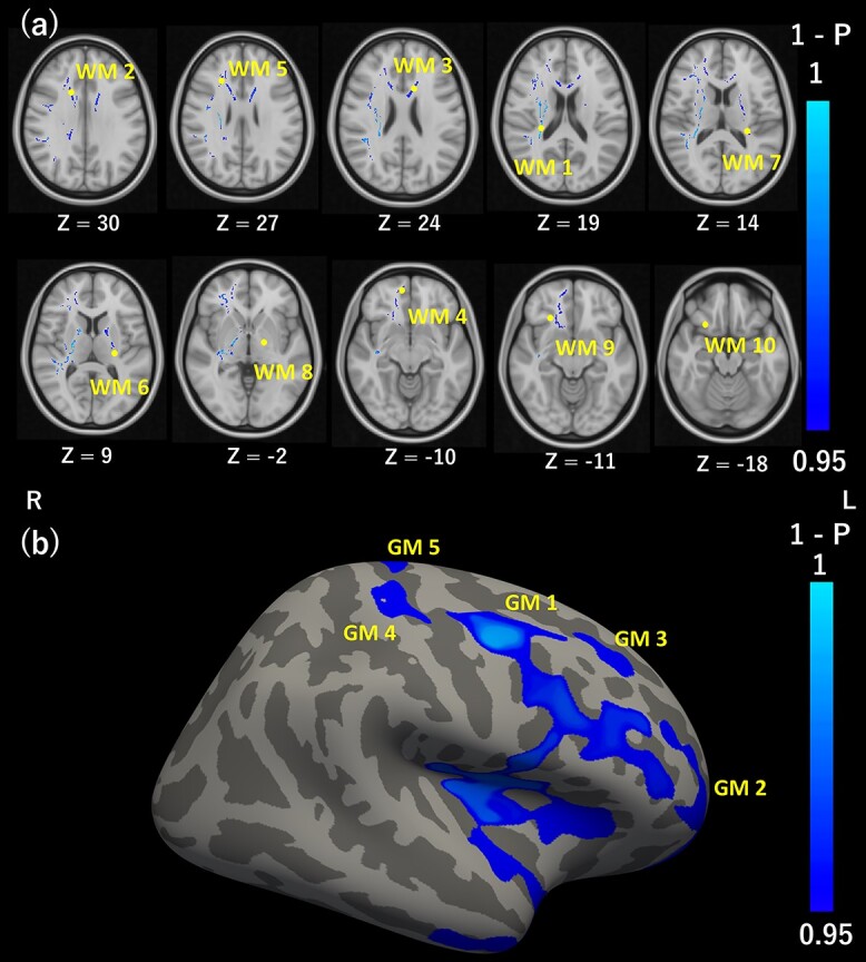
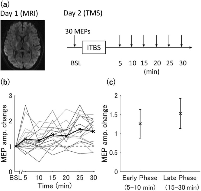
Intermittent theta burst stimulation (iTBS) delivered by transcranial magnetic stimulation (TMS) produces a long-term potentiation-like after-effect useful for investigations of cortical function and of potential therapeutic value. However, the iTBS after-effect over the primary motor cortex (M1) as measured by changes in motor evoked potential (MEP) amplitude exhibits a largely unexplained variability across individuals. Here, we present evidence that individual differences in white matter (WM) and gray matter (GM) microstructural properties revealed by fractional anisotropy (FA) predict the magnitude of the iTBS-induced after-effect over M1. The MEP amplitude change in the early phase (5-10 min post-iTBS) was associated with FA values in WM tracts such as right superior longitudinal fasciculus and corpus callosum. By contrast, the MEP amplitude change in the late phase (15-30 min post-iTBS) was associated with FA in GM, primarily in right frontal cortex. These results suggest that the microstructural properties of regions connected directly or indirectly to the target region (M1) are crucial determinants of the iTBS after-effect. FA values indicative of these microstructural differences can predict the potential effectiveness of repetitive TMS for both investigational use and clinical application.

 求助内容:
求助内容: 应助结果提醒方式:


