E Valas Teuma, Frank A Bucci, Raman Bedi, Gary Gray, Mark Packer
{"title":"飞秒激光部分厚度桡骨和弓形切口在猪眼中的准确性和安全性。","authors":"E Valas Teuma, Frank A Bucci, Raman Bedi, Gary Gray, Mark Packer","doi":"10.1186/s40662-021-00268-w","DOIUrl":null,"url":null,"abstract":"<p><strong>Background: </strong>To evaluate the accuracy and safety of micro radial and arcuate keratotomy incisions constructed by a femtosecond laser system with a curved contact patient interface in porcine eyes.</p><p><strong>Methods: </strong>Partial thickness micro radial and arcuate keratotomy incisions were constructed in porcine eyes with a femtosecond laser system and evaluated for precision of depth, quality, and consistency. Optical coherence tomography was used to determine the accuracy and precision of incision depth. Corneal endothelial safety was assessed by a fluorescent live/dead cell viability assay to demonstrate laser-induced endothelial cell loss. Quality was evaluated by ease of opening and examination of interfaces.</p><p><strong>Results: </strong>In two micro radial incision groups, intended incision depths of 50% and 80% resulted in mean achieved depths of 50.01% and 77.69%, respectively. In three arcuate incision groups, intended incision depths of 80%, 600 μm or 100 μm residual uncut bed thickness resulted in mean achieved depths of 80.16%, 603.03 μm and residual bed of 115 μm, respectively. No loss of endothelial cell density occurred when the residual corneal bed was maintained at a minimum of 85-116 µm. The incisions were easy to open, and interfaces were smooth.</p><p><strong>Conclusions: </strong>A femtosecond laser system with curved contact interface created precise and reproducible micro radial and arcuate keratotomy incisions. Accuracy and precision of the incision depth and preservation of endothelial cell density demonstrated the effectiveness and safety of the system.</p>","PeriodicalId":520624,"journal":{"name":"Eye and vision (London, England)","volume":" ","pages":"41"},"PeriodicalIF":0.0000,"publicationDate":"2021-12-01","publicationTypes":"Journal Article","fieldsOfStudy":null,"isOpenAccess":false,"openAccessPdf":"https://www.ncbi.nlm.nih.gov/pmc/articles/PMC8638553/pdf/","citationCount":"0","resultStr":"{\"title\":\"Accuracy and safety of partial thickness femtosecond laser radial and arcuate keratotomy incisions in porcine eyes.\",\"authors\":\"E Valas Teuma, Frank A Bucci, Raman Bedi, Gary Gray, Mark Packer\",\"doi\":\"10.1186/s40662-021-00268-w\",\"DOIUrl\":null,\"url\":null,\"abstract\":\"<p><strong>Background: </strong>To evaluate the accuracy and safety of micro radial and arcuate keratotomy incisions constructed by a femtosecond laser system with a curved contact patient interface in porcine eyes.</p><p><strong>Methods: </strong>Partial thickness micro radial and arcuate keratotomy incisions were constructed in porcine eyes with a femtosecond laser system and evaluated for precision of depth, quality, and consistency. Optical coherence tomography was used to determine the accuracy and precision of incision depth. Corneal endothelial safety was assessed by a fluorescent live/dead cell viability assay to demonstrate laser-induced endothelial cell loss. Quality was evaluated by ease of opening and examination of interfaces.</p><p><strong>Results: </strong>In two micro radial incision groups, intended incision depths of 50% and 80% resulted in mean achieved depths of 50.01% and 77.69%, respectively. In three arcuate incision groups, intended incision depths of 80%, 600 μm or 100 μm residual uncut bed thickness resulted in mean achieved depths of 80.16%, 603.03 μm and residual bed of 115 μm, respectively. No loss of endothelial cell density occurred when the residual corneal bed was maintained at a minimum of 85-116 µm. The incisions were easy to open, and interfaces were smooth.</p><p><strong>Conclusions: </strong>A femtosecond laser system with curved contact interface created precise and reproducible micro radial and arcuate keratotomy incisions. Accuracy and precision of the incision depth and preservation of endothelial cell density demonstrated the effectiveness and safety of the system.</p>\",\"PeriodicalId\":520624,\"journal\":{\"name\":\"Eye and vision (London, England)\",\"volume\":\" \",\"pages\":\"41\"},\"PeriodicalIF\":0.0000,\"publicationDate\":\"2021-12-01\",\"publicationTypes\":\"Journal Article\",\"fieldsOfStudy\":null,\"isOpenAccess\":false,\"openAccessPdf\":\"https://www.ncbi.nlm.nih.gov/pmc/articles/PMC8638553/pdf/\",\"citationCount\":\"0\",\"resultStr\":null,\"platform\":\"Semanticscholar\",\"paperid\":null,\"PeriodicalName\":\"Eye and vision (London, England)\",\"FirstCategoryId\":\"3\",\"ListUrlMain\":\"https://doi.org/10.1186/s40662-021-00268-w\",\"RegionNum\":0,\"RegionCategory\":null,\"ArticlePicture\":[],\"TitleCN\":null,\"AbstractTextCN\":null,\"PMCID\":null,\"EPubDate\":\"\",\"PubModel\":\"\",\"JCR\":\"\",\"JCRName\":\"\",\"Score\":null,\"Total\":0}","platform":"Semanticscholar","paperid":null,"PeriodicalName":"Eye and vision (London, England)","FirstCategoryId":"3","ListUrlMain":"https://doi.org/10.1186/s40662-021-00268-w","RegionNum":0,"RegionCategory":null,"ArticlePicture":[],"TitleCN":null,"AbstractTextCN":null,"PMCID":null,"EPubDate":"","PubModel":"","JCR":"","JCRName":"","Score":null,"Total":0}
Accuracy and safety of partial thickness femtosecond laser radial and arcuate keratotomy incisions in porcine eyes.
Background: To evaluate the accuracy and safety of micro radial and arcuate keratotomy incisions constructed by a femtosecond laser system with a curved contact patient interface in porcine eyes.
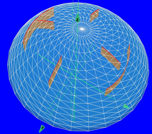
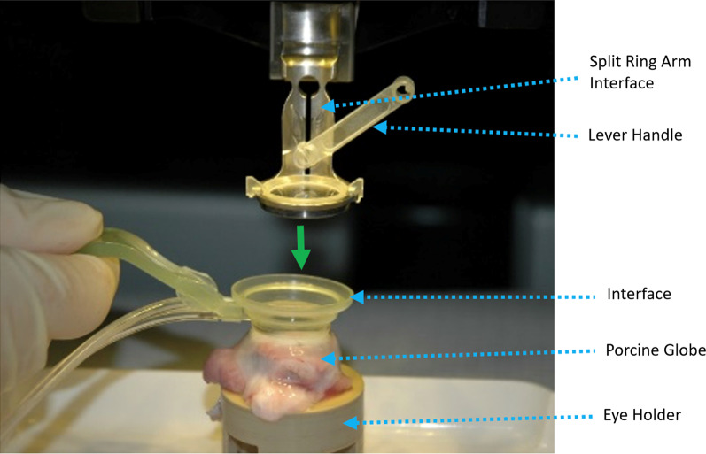
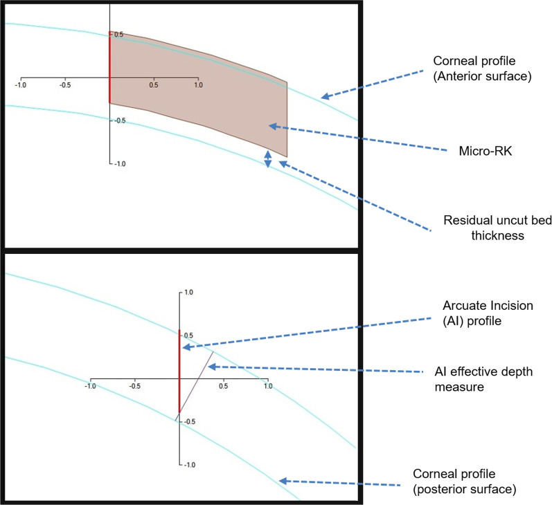
Methods: Partial thickness micro radial and arcuate keratotomy incisions were constructed in porcine eyes with a femtosecond laser system and evaluated for precision of depth, quality, and consistency. Optical coherence tomography was used to determine the accuracy and precision of incision depth. Corneal endothelial safety was assessed by a fluorescent live/dead cell viability assay to demonstrate laser-induced endothelial cell loss. Quality was evaluated by ease of opening and examination of interfaces.
Results: In two micro radial incision groups, intended incision depths of 50% and 80% resulted in mean achieved depths of 50.01% and 77.69%, respectively. In three arcuate incision groups, intended incision depths of 80%, 600 μm or 100 μm residual uncut bed thickness resulted in mean achieved depths of 80.16%, 603.03 μm and residual bed of 115 μm, respectively. No loss of endothelial cell density occurred when the residual corneal bed was maintained at a minimum of 85-116 µm. The incisions were easy to open, and interfaces were smooth.
Conclusions: A femtosecond laser system with curved contact interface created precise and reproducible micro radial and arcuate keratotomy incisions. Accuracy and precision of the incision depth and preservation of endothelial cell density demonstrated the effectiveness and safety of the system.

 求助内容:
求助内容: 应助结果提醒方式:


