Michał Kunc, Alexandra Kamieniecki, Grzegorz Walczak, Tomasz Nowicki, Bartosz Wasąg, Bogusław Mikaszewski, Dominik Stodulski, Wojciech Biernat
{"title":"涎腺内胸腺癌1例报告及文献复习。","authors":"Michał Kunc, Alexandra Kamieniecki, Grzegorz Walczak, Tomasz Nowicki, Bartosz Wasąg, Bogusław Mikaszewski, Dominik Stodulski, Wojciech Biernat","doi":"10.1007/s12105-021-01394-6","DOIUrl":null,"url":null,"abstract":"<p><p>Ectopic thymic carcinomas are rarely diagnosed in the thyroid gland, let alone in extrathyroid tissues. In the currently available literature, only five cases of extrathyroidal malignancies with thymic differentiation have been reported as arising in the major salivary glands. A 69-year-old female presented with a slow-growing palpable mass in the left parotid gland. Fine needle aspiration biopsy suggested metastatic cancer, whereas core needle biopsy revealed high-grade squamous cell carcinoma. The patient underwent left radical parotidectomy with selective ipsilateral lymph node dissection and subsequent radiation therapy. The surgical specimen was taken for histopathological examination. Microscopically, the tumor resembled thymic carcinoma. It was composed of large nests of squamoid cells with smooth contours, focally with a syncytial growth pattern, and accompanied by abundant lymphocytes with reactive lymphoid follicles. This appearance resembled a micronodular thymic carcinoma with lymphoid hyperplasia. Moreover, the tumor displayed expression of squamous markers (p40 and p63) and markers of thymic carcinoma (CD5 and CD117). Therefore, the final diagnosis of intrasalivary thymic carcinoma was rendered. The molecular analysis including next-generation sequencing demonstrated no variants of the strong, potential, or unknown clinical significance. The patient remains disease-free at 1-year follow-up. In the current case, we comprehensively present a clinical, microscopic, molecular, and radiological picture of CD5-positive squamous cell carcinoma of the parotid. We postulate that similar cases should be designated as intrasalivary thymic carcinoma analogically to similar thyroid tumors. Our case and the limited literature data indicate they should be distinguished from conventional squamous cell carcinoma of major salivary glands due to their rather favorable prognosis.</p>","PeriodicalId":520636,"journal":{"name":"Head and neck pathology","volume":" ","pages":"857-864"},"PeriodicalIF":0.0000,"publicationDate":"2022-09-01","publicationTypes":"Journal Article","fieldsOfStudy":null,"isOpenAccess":false,"openAccessPdf":"https://www.ncbi.nlm.nih.gov/pmc/articles/PMC9424426/pdf/","citationCount":"4","resultStr":"{\"title\":\"Intrasalivary Thymic Carcinoma: A Case Report and Literature Review.\",\"authors\":\"Michał Kunc, Alexandra Kamieniecki, Grzegorz Walczak, Tomasz Nowicki, Bartosz Wasąg, Bogusław Mikaszewski, Dominik Stodulski, Wojciech Biernat\",\"doi\":\"10.1007/s12105-021-01394-6\",\"DOIUrl\":null,\"url\":null,\"abstract\":\"<p><p>Ectopic thymic carcinomas are rarely diagnosed in the thyroid gland, let alone in extrathyroid tissues. In the currently available literature, only five cases of extrathyroidal malignancies with thymic differentiation have been reported as arising in the major salivary glands. A 69-year-old female presented with a slow-growing palpable mass in the left parotid gland. Fine needle aspiration biopsy suggested metastatic cancer, whereas core needle biopsy revealed high-grade squamous cell carcinoma. The patient underwent left radical parotidectomy with selective ipsilateral lymph node dissection and subsequent radiation therapy. The surgical specimen was taken for histopathological examination. Microscopically, the tumor resembled thymic carcinoma. It was composed of large nests of squamoid cells with smooth contours, focally with a syncytial growth pattern, and accompanied by abundant lymphocytes with reactive lymphoid follicles. This appearance resembled a micronodular thymic carcinoma with lymphoid hyperplasia. Moreover, the tumor displayed expression of squamous markers (p40 and p63) and markers of thymic carcinoma (CD5 and CD117). Therefore, the final diagnosis of intrasalivary thymic carcinoma was rendered. The molecular analysis including next-generation sequencing demonstrated no variants of the strong, potential, or unknown clinical significance. The patient remains disease-free at 1-year follow-up. In the current case, we comprehensively present a clinical, microscopic, molecular, and radiological picture of CD5-positive squamous cell carcinoma of the parotid. We postulate that similar cases should be designated as intrasalivary thymic carcinoma analogically to similar thyroid tumors. Our case and the limited literature data indicate they should be distinguished from conventional squamous cell carcinoma of major salivary glands due to their rather favorable prognosis.</p>\",\"PeriodicalId\":520636,\"journal\":{\"name\":\"Head and neck pathology\",\"volume\":\" \",\"pages\":\"857-864\"},\"PeriodicalIF\":0.0000,\"publicationDate\":\"2022-09-01\",\"publicationTypes\":\"Journal Article\",\"fieldsOfStudy\":null,\"isOpenAccess\":false,\"openAccessPdf\":\"https://www.ncbi.nlm.nih.gov/pmc/articles/PMC9424426/pdf/\",\"citationCount\":\"4\",\"resultStr\":null,\"platform\":\"Semanticscholar\",\"paperid\":null,\"PeriodicalName\":\"Head and neck pathology\",\"FirstCategoryId\":\"1085\",\"ListUrlMain\":\"https://doi.org/10.1007/s12105-021-01394-6\",\"RegionNum\":0,\"RegionCategory\":null,\"ArticlePicture\":[],\"TitleCN\":null,\"AbstractTextCN\":null,\"PMCID\":null,\"EPubDate\":\"2021/11/22 0:00:00\",\"PubModel\":\"Epub\",\"JCR\":\"\",\"JCRName\":\"\",\"Score\":null,\"Total\":0}","platform":"Semanticscholar","paperid":null,"PeriodicalName":"Head and neck pathology","FirstCategoryId":"1085","ListUrlMain":"https://doi.org/10.1007/s12105-021-01394-6","RegionNum":0,"RegionCategory":null,"ArticlePicture":[],"TitleCN":null,"AbstractTextCN":null,"PMCID":null,"EPubDate":"2021/11/22 0:00:00","PubModel":"Epub","JCR":"","JCRName":"","Score":null,"Total":0}
Intrasalivary Thymic Carcinoma: A Case Report and Literature Review.
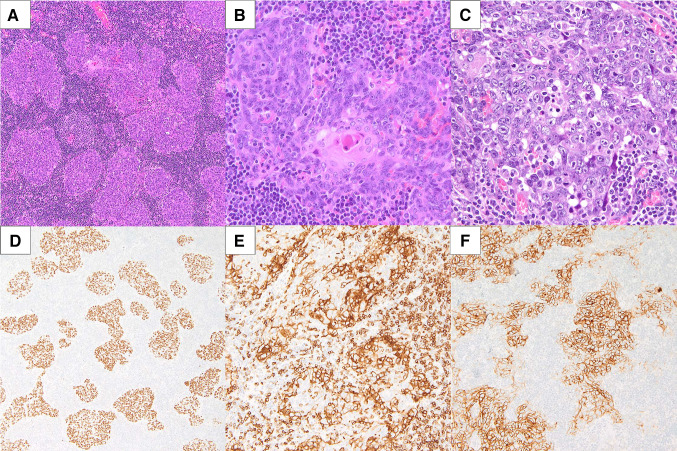
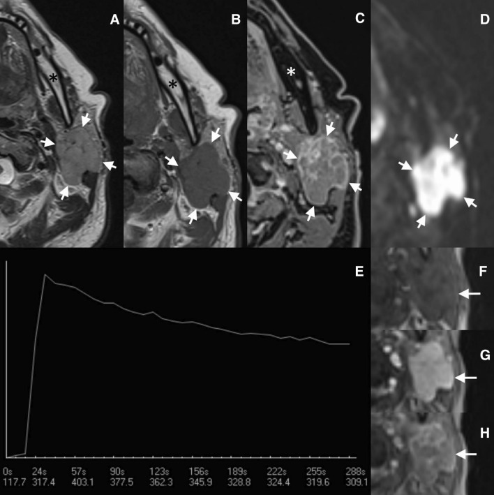
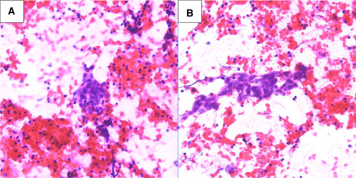
Ectopic thymic carcinomas are rarely diagnosed in the thyroid gland, let alone in extrathyroid tissues. In the currently available literature, only five cases of extrathyroidal malignancies with thymic differentiation have been reported as arising in the major salivary glands. A 69-year-old female presented with a slow-growing palpable mass in the left parotid gland. Fine needle aspiration biopsy suggested metastatic cancer, whereas core needle biopsy revealed high-grade squamous cell carcinoma. The patient underwent left radical parotidectomy with selective ipsilateral lymph node dissection and subsequent radiation therapy. The surgical specimen was taken for histopathological examination. Microscopically, the tumor resembled thymic carcinoma. It was composed of large nests of squamoid cells with smooth contours, focally with a syncytial growth pattern, and accompanied by abundant lymphocytes with reactive lymphoid follicles. This appearance resembled a micronodular thymic carcinoma with lymphoid hyperplasia. Moreover, the tumor displayed expression of squamous markers (p40 and p63) and markers of thymic carcinoma (CD5 and CD117). Therefore, the final diagnosis of intrasalivary thymic carcinoma was rendered. The molecular analysis including next-generation sequencing demonstrated no variants of the strong, potential, or unknown clinical significance. The patient remains disease-free at 1-year follow-up. In the current case, we comprehensively present a clinical, microscopic, molecular, and radiological picture of CD5-positive squamous cell carcinoma of the parotid. We postulate that similar cases should be designated as intrasalivary thymic carcinoma analogically to similar thyroid tumors. Our case and the limited literature data indicate they should be distinguished from conventional squamous cell carcinoma of major salivary glands due to their rather favorable prognosis.

 求助内容:
求助内容: 应助结果提醒方式:


