{"title":"鞍上黑素细胞瘤伴轻脑膜播种:组织学上良性肿瘤的侵袭性临床病程。","authors":"Imen Maaloul, Marwa Moussaoui, Ameni Salah, Wiem Feki, Hela Fourati, Nadia Charfi, Zeineb Mnif","doi":"10.1155/2021/7306432","DOIUrl":null,"url":null,"abstract":"<p><strong>Introduction: </strong>Meningeal melanocytoma (MM) is a very rare neuroectodermal neoplasm arising from the leptomeninges. Primary suprasellar melanocytomas are exceedingly rare, with only a handful of cases reported. The systemic spread of a nontransformed meningeal melanocytoma is an unusual occurrence. Herein, we report the first case of a primary sellar melanocytoma with cerebral and spinal meningeal seeding. <i>Case Report</i>. A 30-year-old male with no previous medical history presented to the endocrinology department with a loss of body hair. The endocrine workup concluded with isolated hypogonadotropic hypogonadism. The Magnetic Resonance Imaging (MRI) of the brain and sella revealed a large suprasellar mass continuous with the infundibulum of the pituitary gland. It was heterogeneously hyperintense on T1-, T2-, and FLAIR-weighted images and was enhanced with contrast, along with cerebral and spinal leptomeningeal spread. The patient was referred to the neurosurgery department, and a lumbar spine biopsy was indicated. The histopathological examination was suggestive of a grade I meningeal pigmented melanocytoma.</p><p><strong>Conclusion: </strong>Thus, primary sellar melanocytomas with leptomeningeal spread are an extremely rare phenomenon. Metastatic malignant melanoma should be ruled out. Being aware of differential diagnosis and the unusual behavior of meningeal melanocytoma will be necessary to manage the patient appropriately. Complete tumor resection is the best treatment whenever possible, and radiotherapy should be considered in case of unresectability or partial resection.</p>","PeriodicalId":30326,"journal":{"name":"Case Reports in Radiology","volume":" ","pages":"7306432"},"PeriodicalIF":0.0000,"publicationDate":"2021-10-11","publicationTypes":"Journal Article","fieldsOfStudy":null,"isOpenAccess":false,"openAccessPdf":"https://www.ncbi.nlm.nih.gov/pmc/articles/PMC8523264/pdf/","citationCount":"1","resultStr":"{\"title\":\"Suprasellar Melanocytoma with Leptomeningeal Seeding: An Aggressive Clinical Course for a Histologically Benign Tumor.\",\"authors\":\"Imen Maaloul, Marwa Moussaoui, Ameni Salah, Wiem Feki, Hela Fourati, Nadia Charfi, Zeineb Mnif\",\"doi\":\"10.1155/2021/7306432\",\"DOIUrl\":null,\"url\":null,\"abstract\":\"<p><strong>Introduction: </strong>Meningeal melanocytoma (MM) is a very rare neuroectodermal neoplasm arising from the leptomeninges. Primary suprasellar melanocytomas are exceedingly rare, with only a handful of cases reported. The systemic spread of a nontransformed meningeal melanocytoma is an unusual occurrence. Herein, we report the first case of a primary sellar melanocytoma with cerebral and spinal meningeal seeding. <i>Case Report</i>. A 30-year-old male with no previous medical history presented to the endocrinology department with a loss of body hair. The endocrine workup concluded with isolated hypogonadotropic hypogonadism. The Magnetic Resonance Imaging (MRI) of the brain and sella revealed a large suprasellar mass continuous with the infundibulum of the pituitary gland. It was heterogeneously hyperintense on T1-, T2-, and FLAIR-weighted images and was enhanced with contrast, along with cerebral and spinal leptomeningeal spread. The patient was referred to the neurosurgery department, and a lumbar spine biopsy was indicated. The histopathological examination was suggestive of a grade I meningeal pigmented melanocytoma.</p><p><strong>Conclusion: </strong>Thus, primary sellar melanocytomas with leptomeningeal spread are an extremely rare phenomenon. Metastatic malignant melanoma should be ruled out. Being aware of differential diagnosis and the unusual behavior of meningeal melanocytoma will be necessary to manage the patient appropriately. Complete tumor resection is the best treatment whenever possible, and radiotherapy should be considered in case of unresectability or partial resection.</p>\",\"PeriodicalId\":30326,\"journal\":{\"name\":\"Case Reports in Radiology\",\"volume\":\" \",\"pages\":\"7306432\"},\"PeriodicalIF\":0.0000,\"publicationDate\":\"2021-10-11\",\"publicationTypes\":\"Journal Article\",\"fieldsOfStudy\":null,\"isOpenAccess\":false,\"openAccessPdf\":\"https://www.ncbi.nlm.nih.gov/pmc/articles/PMC8523264/pdf/\",\"citationCount\":\"1\",\"resultStr\":null,\"platform\":\"Semanticscholar\",\"paperid\":null,\"PeriodicalName\":\"Case Reports in Radiology\",\"FirstCategoryId\":\"1085\",\"ListUrlMain\":\"https://doi.org/10.1155/2021/7306432\",\"RegionNum\":0,\"RegionCategory\":null,\"ArticlePicture\":[],\"TitleCN\":null,\"AbstractTextCN\":null,\"PMCID\":null,\"EPubDate\":\"2021/1/1 0:00:00\",\"PubModel\":\"eCollection\",\"JCR\":\"\",\"JCRName\":\"\",\"Score\":null,\"Total\":0}","platform":"Semanticscholar","paperid":null,"PeriodicalName":"Case Reports in Radiology","FirstCategoryId":"1085","ListUrlMain":"https://doi.org/10.1155/2021/7306432","RegionNum":0,"RegionCategory":null,"ArticlePicture":[],"TitleCN":null,"AbstractTextCN":null,"PMCID":null,"EPubDate":"2021/1/1 0:00:00","PubModel":"eCollection","JCR":"","JCRName":"","Score":null,"Total":0}
Suprasellar Melanocytoma with Leptomeningeal Seeding: An Aggressive Clinical Course for a Histologically Benign Tumor.
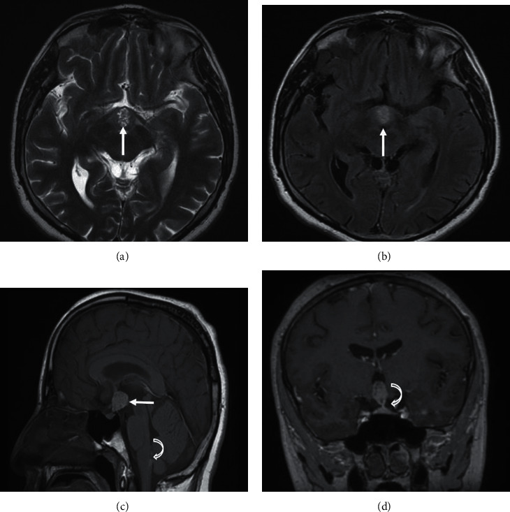
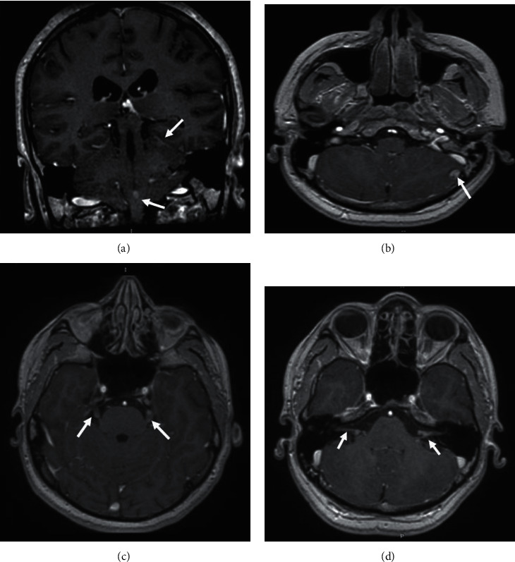
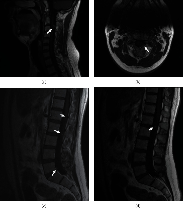
Introduction: Meningeal melanocytoma (MM) is a very rare neuroectodermal neoplasm arising from the leptomeninges. Primary suprasellar melanocytomas are exceedingly rare, with only a handful of cases reported. The systemic spread of a nontransformed meningeal melanocytoma is an unusual occurrence. Herein, we report the first case of a primary sellar melanocytoma with cerebral and spinal meningeal seeding. Case Report. A 30-year-old male with no previous medical history presented to the endocrinology department with a loss of body hair. The endocrine workup concluded with isolated hypogonadotropic hypogonadism. The Magnetic Resonance Imaging (MRI) of the brain and sella revealed a large suprasellar mass continuous with the infundibulum of the pituitary gland. It was heterogeneously hyperintense on T1-, T2-, and FLAIR-weighted images and was enhanced with contrast, along with cerebral and spinal leptomeningeal spread. The patient was referred to the neurosurgery department, and a lumbar spine biopsy was indicated. The histopathological examination was suggestive of a grade I meningeal pigmented melanocytoma.
Conclusion: Thus, primary sellar melanocytomas with leptomeningeal spread are an extremely rare phenomenon. Metastatic malignant melanoma should be ruled out. Being aware of differential diagnosis and the unusual behavior of meningeal melanocytoma will be necessary to manage the patient appropriately. Complete tumor resection is the best treatment whenever possible, and radiotherapy should be considered in case of unresectability or partial resection.

 求助内容:
求助内容: 应助结果提醒方式:


