Jens Ingwersen, Jonas Graf, Julia Kluge, Margit Weise, Michael Dietrich, John-Ih Lee, Jens Harmel, Hans-Peter Hartung, Tobias Ruck, Sven G Meuth, Philipp Albrecht, Orhan Aktas, Marius Ringelstein
{"title":"慢性炎症性脱髓鞘性多神经病变的中枢神经系统参与:光学相干断层扫描的细微视网膜变化。","authors":"Jens Ingwersen, Jonas Graf, Julia Kluge, Margit Weise, Michael Dietrich, John-Ih Lee, Jens Harmel, Hans-Peter Hartung, Tobias Ruck, Sven G Meuth, Philipp Albrecht, Orhan Aktas, Marius Ringelstein","doi":"10.1212/NXI.0000000000001099","DOIUrl":null,"url":null,"abstract":"<p><strong>Background and objectives: </strong>Chronic inflammatory demyelinating polyneuropathy (CIDP) is an autoimmune disease primarily affecting the peripheral nervous system. However, several noncontrolled studies have suggested concomitant inflammatory CNS demyelination similar to multiple sclerosis. The aim of this study was to investigate an involvement of the visual pathway in patients with CIDP.</p><p><strong>Methods: </strong>In this prospective cross-sectional study, we used high-resolution spectral-domain optical coherence tomography to compare the thickness of the peripapillary retinal nerve fiber layer and the deeper macular retinal layers as well as the total macular volume (TMV) in 22 patients with CIDP and 22 age-matched and sex-matched healthy control (HC) individuals. Retinal layers were semiautomatically segmented by the provided software and were correlated with clinical measures and nerve conduction studies.</p><p><strong>Results: </strong>In patients with CIDP compared with healthy age-matched and sex-matched controls, we found slight but significant volume reductions of the ganglion cell/inner plexiform layer complex (CIDP 1.86 vs HC 1.95 mm<sup>3</sup>, <i>p</i> = 0.015), the retinal pigment epithelium (CIDP 0.38 vs HC 0.40 mm<sup>3</sup>, <i>p</i> = 0.02), and the TMV (CIDP 8.48 vs HC 8.75 mm<sup>3</sup>, <i>p</i> = 0.018). The ganglion cell layer volume and motor nerve conduction velocity were positively associated (B = 0.002, <i>p</i> = 0.02).</p><p><strong>Discussion: </strong>Our data reveal subtle retinal neurodegeneration in patients with CIDP, providing evidence for visual pathway involvement, detectable by OCT. The results need corroboration in independent, larger cohorts.</p>","PeriodicalId":520720,"journal":{"name":"Neurology(R) neuroimmunology & neuroinflammation","volume":" ","pages":""},"PeriodicalIF":0.0000,"publicationDate":"2021-10-19","publicationTypes":"Journal Article","fieldsOfStudy":null,"isOpenAccess":false,"openAccessPdf":"https://ftp.ncbi.nlm.nih.gov/pub/pmc/oa_pdf/e4/c1/NEURIMMINFL2021039086.PMC8529418.pdf","citationCount":"2","resultStr":"{\"title\":\"CNS Involvement in Chronic Inflammatory Demyelinating Polyneuropathy: Subtle Retinal Changes in Optical Coherence Tomography.\",\"authors\":\"Jens Ingwersen, Jonas Graf, Julia Kluge, Margit Weise, Michael Dietrich, John-Ih Lee, Jens Harmel, Hans-Peter Hartung, Tobias Ruck, Sven G Meuth, Philipp Albrecht, Orhan Aktas, Marius Ringelstein\",\"doi\":\"10.1212/NXI.0000000000001099\",\"DOIUrl\":null,\"url\":null,\"abstract\":\"<p><strong>Background and objectives: </strong>Chronic inflammatory demyelinating polyneuropathy (CIDP) is an autoimmune disease primarily affecting the peripheral nervous system. However, several noncontrolled studies have suggested concomitant inflammatory CNS demyelination similar to multiple sclerosis. The aim of this study was to investigate an involvement of the visual pathway in patients with CIDP.</p><p><strong>Methods: </strong>In this prospective cross-sectional study, we used high-resolution spectral-domain optical coherence tomography to compare the thickness of the peripapillary retinal nerve fiber layer and the deeper macular retinal layers as well as the total macular volume (TMV) in 22 patients with CIDP and 22 age-matched and sex-matched healthy control (HC) individuals. Retinal layers were semiautomatically segmented by the provided software and were correlated with clinical measures and nerve conduction studies.</p><p><strong>Results: </strong>In patients with CIDP compared with healthy age-matched and sex-matched controls, we found slight but significant volume reductions of the ganglion cell/inner plexiform layer complex (CIDP 1.86 vs HC 1.95 mm<sup>3</sup>, <i>p</i> = 0.015), the retinal pigment epithelium (CIDP 0.38 vs HC 0.40 mm<sup>3</sup>, <i>p</i> = 0.02), and the TMV (CIDP 8.48 vs HC 8.75 mm<sup>3</sup>, <i>p</i> = 0.018). The ganglion cell layer volume and motor nerve conduction velocity were positively associated (B = 0.002, <i>p</i> = 0.02).</p><p><strong>Discussion: </strong>Our data reveal subtle retinal neurodegeneration in patients with CIDP, providing evidence for visual pathway involvement, detectable by OCT. The results need corroboration in independent, larger cohorts.</p>\",\"PeriodicalId\":520720,\"journal\":{\"name\":\"Neurology(R) neuroimmunology & neuroinflammation\",\"volume\":\" \",\"pages\":\"\"},\"PeriodicalIF\":0.0000,\"publicationDate\":\"2021-10-19\",\"publicationTypes\":\"Journal Article\",\"fieldsOfStudy\":null,\"isOpenAccess\":false,\"openAccessPdf\":\"https://ftp.ncbi.nlm.nih.gov/pub/pmc/oa_pdf/e4/c1/NEURIMMINFL2021039086.PMC8529418.pdf\",\"citationCount\":\"2\",\"resultStr\":null,\"platform\":\"Semanticscholar\",\"paperid\":null,\"PeriodicalName\":\"Neurology(R) neuroimmunology & neuroinflammation\",\"FirstCategoryId\":\"3\",\"ListUrlMain\":\"https://doi.org/10.1212/NXI.0000000000001099\",\"RegionNum\":0,\"RegionCategory\":null,\"ArticlePicture\":[],\"TitleCN\":null,\"AbstractTextCN\":null,\"PMCID\":null,\"EPubDate\":\"2022/1/1 0:00:00\",\"PubModel\":\"Print\",\"JCR\":\"\",\"JCRName\":\"\",\"Score\":null,\"Total\":0}","platform":"Semanticscholar","paperid":null,"PeriodicalName":"Neurology(R) neuroimmunology & neuroinflammation","FirstCategoryId":"3","ListUrlMain":"https://doi.org/10.1212/NXI.0000000000001099","RegionNum":0,"RegionCategory":null,"ArticlePicture":[],"TitleCN":null,"AbstractTextCN":null,"PMCID":null,"EPubDate":"2022/1/1 0:00:00","PubModel":"Print","JCR":"","JCRName":"","Score":null,"Total":0}
引用次数: 2
摘要
背景和目的:慢性炎症性脱髓鞘性多神经病变(CIDP)是一种主要影响周围神经系统的自身免疫性疾病。然而,一些非对照研究表明,伴有炎症性中枢神经系统脱髓鞘类似于多发性硬化症。本研究的目的是探讨CIDP患者的视觉通路的参与。方法:在这项前瞻性横断面研究中,我们使用高分辨率光谱域光学相干断层扫描比较了22例CIDP患者和22例年龄匹配和性别匹配的健康对照(HC)患者的乳头周围视网膜神经纤维层和更深黄斑视网膜层的厚度以及黄斑总体积(TMV)。通过提供的软件半自动分割视网膜层,并与临床测量和神经传导研究相关联。结果:与年龄匹配和性别匹配的健康对照组相比,CIDP患者的神经节细胞/内丛状层复合体(CIDP 1.86 vs HC 1.95 mm3, p = 0.015)、视网膜色素上皮(CIDP 0.38 vs HC 0.40 mm3, p = 0.02)和TMV (CIDP 8.48 vs HC 8.75 mm3, p = 0.018)体积轻微但显著减少。神经节细胞层体积与运动神经传导速度呈正相关(B = 0.002, p = 0.02)。讨论:我们的数据揭示了CIDP患者的细微视网膜神经退行性变,提供了视觉通路受累的证据,可以通过oct检测到。结果需要在独立的、更大的队列中得到证实。
CNS Involvement in Chronic Inflammatory Demyelinating Polyneuropathy: Subtle Retinal Changes in Optical Coherence Tomography.
Background and objectives: Chronic inflammatory demyelinating polyneuropathy (CIDP) is an autoimmune disease primarily affecting the peripheral nervous system. However, several noncontrolled studies have suggested concomitant inflammatory CNS demyelination similar to multiple sclerosis. The aim of this study was to investigate an involvement of the visual pathway in patients with CIDP.
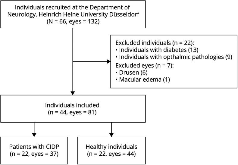
Methods: In this prospective cross-sectional study, we used high-resolution spectral-domain optical coherence tomography to compare the thickness of the peripapillary retinal nerve fiber layer and the deeper macular retinal layers as well as the total macular volume (TMV) in 22 patients with CIDP and 22 age-matched and sex-matched healthy control (HC) individuals. Retinal layers were semiautomatically segmented by the provided software and were correlated with clinical measures and nerve conduction studies.
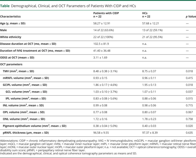
Results: In patients with CIDP compared with healthy age-matched and sex-matched controls, we found slight but significant volume reductions of the ganglion cell/inner plexiform layer complex (CIDP 1.86 vs HC 1.95 mm3, p = 0.015), the retinal pigment epithelium (CIDP 0.38 vs HC 0.40 mm3, p = 0.02), and the TMV (CIDP 8.48 vs HC 8.75 mm3, p = 0.018). The ganglion cell layer volume and motor nerve conduction velocity were positively associated (B = 0.002, p = 0.02).
Discussion: Our data reveal subtle retinal neurodegeneration in patients with CIDP, providing evidence for visual pathway involvement, detectable by OCT. The results need corroboration in independent, larger cohorts.

 求助内容:
求助内容: 应助结果提醒方式:


