Raafat Mohyeldeen Abdelrahman Abdallah, Ahmed Mohamed Kamal Elshafei, Heba Radi AttaAllah
{"title":"应用前段光学相干断层扫描评价植入穿孔泪点塞。","authors":"Raafat Mohyeldeen Abdelrahman Abdallah, Ahmed Mohamed Kamal Elshafei, Heba Radi AttaAllah","doi":"10.1186/s40662-021-00259-x","DOIUrl":null,"url":null,"abstract":"<p><strong>Purpose: </strong>Evaluation of the patency and position of perforated lacrimal punctal plugs implanted for treating punctal stenosis together with quantitative assessment of the precorneal tear film using anterior segment optical coherence tomography (AS-OCT).</p><p><strong>Methods: </strong>In a prospective study, the lower punctum of 54 eyes of 29 patients implanted with perforated punctal plugs were examined using AS-OCT during the early postoperative period. Preoperative tear meniscus height (TMH) and tear meniscus area (TMA) were evaluated. Postoperatively, the patency of the plug, its position, TMH and TMA were evaluated, and the results were correlated with postoperative epiphora. Munk scale was used for epiphora grading.</p><p><strong>Results: </strong>Using AS-OCT, 48 (88.9%) plugs were found in proper position while 6 (11.1%) were rotated. The lumen of the plugs was completely patent in 47 (87%) plugs, partially obstructed in 2 (3.7%) plugs and completely occluded in 5 (9.2%) plugs. There was a statistically significant postoperative decrease of TMH and TMA (P < 0.001) and postoperative epiphora Munk score (P < 0.001).</p><p><strong>Conclusion: </strong>AS-OCT is a valuable, reliable, and noninvasive investigative tool that can detect the proper positioning, patency, and contents of the implanted perforated lacrimal punctal plugs in addition to measurement of TMH and TMA. Trial registration ClinicalTrials.gov ID: NCT04624022, https://clinicaltrials.gov/ct2/show/NCT04624022.</p>","PeriodicalId":520624,"journal":{"name":"Eye and vision (London, England)","volume":" ","pages":"36"},"PeriodicalIF":0.0000,"publicationDate":"2021-10-03","publicationTypes":"Journal Article","fieldsOfStudy":null,"isOpenAccess":false,"openAccessPdf":"https://www.ncbi.nlm.nih.gov/pmc/articles/PMC8487482/pdf/","citationCount":"1","resultStr":"{\"title\":\"Evaluation of implanted perforated lacrimal punctal plugs using anterior segment optical coherence tomography.\",\"authors\":\"Raafat Mohyeldeen Abdelrahman Abdallah, Ahmed Mohamed Kamal Elshafei, Heba Radi AttaAllah\",\"doi\":\"10.1186/s40662-021-00259-x\",\"DOIUrl\":null,\"url\":null,\"abstract\":\"<p><strong>Purpose: </strong>Evaluation of the patency and position of perforated lacrimal punctal plugs implanted for treating punctal stenosis together with quantitative assessment of the precorneal tear film using anterior segment optical coherence tomography (AS-OCT).</p><p><strong>Methods: </strong>In a prospective study, the lower punctum of 54 eyes of 29 patients implanted with perforated punctal plugs were examined using AS-OCT during the early postoperative period. Preoperative tear meniscus height (TMH) and tear meniscus area (TMA) were evaluated. Postoperatively, the patency of the plug, its position, TMH and TMA were evaluated, and the results were correlated with postoperative epiphora. Munk scale was used for epiphora grading.</p><p><strong>Results: </strong>Using AS-OCT, 48 (88.9%) plugs were found in proper position while 6 (11.1%) were rotated. The lumen of the plugs was completely patent in 47 (87%) plugs, partially obstructed in 2 (3.7%) plugs and completely occluded in 5 (9.2%) plugs. There was a statistically significant postoperative decrease of TMH and TMA (P < 0.001) and postoperative epiphora Munk score (P < 0.001).</p><p><strong>Conclusion: </strong>AS-OCT is a valuable, reliable, and noninvasive investigative tool that can detect the proper positioning, patency, and contents of the implanted perforated lacrimal punctal plugs in addition to measurement of TMH and TMA. Trial registration ClinicalTrials.gov ID: NCT04624022, https://clinicaltrials.gov/ct2/show/NCT04624022.</p>\",\"PeriodicalId\":520624,\"journal\":{\"name\":\"Eye and vision (London, England)\",\"volume\":\" \",\"pages\":\"36\"},\"PeriodicalIF\":0.0000,\"publicationDate\":\"2021-10-03\",\"publicationTypes\":\"Journal Article\",\"fieldsOfStudy\":null,\"isOpenAccess\":false,\"openAccessPdf\":\"https://www.ncbi.nlm.nih.gov/pmc/articles/PMC8487482/pdf/\",\"citationCount\":\"1\",\"resultStr\":null,\"platform\":\"Semanticscholar\",\"paperid\":null,\"PeriodicalName\":\"Eye and vision (London, England)\",\"FirstCategoryId\":\"3\",\"ListUrlMain\":\"https://doi.org/10.1186/s40662-021-00259-x\",\"RegionNum\":0,\"RegionCategory\":null,\"ArticlePicture\":[],\"TitleCN\":null,\"AbstractTextCN\":null,\"PMCID\":null,\"EPubDate\":\"\",\"PubModel\":\"\",\"JCR\":\"\",\"JCRName\":\"\",\"Score\":null,\"Total\":0}","platform":"Semanticscholar","paperid":null,"PeriodicalName":"Eye and vision (London, England)","FirstCategoryId":"3","ListUrlMain":"https://doi.org/10.1186/s40662-021-00259-x","RegionNum":0,"RegionCategory":null,"ArticlePicture":[],"TitleCN":null,"AbstractTextCN":null,"PMCID":null,"EPubDate":"","PubModel":"","JCR":"","JCRName":"","Score":null,"Total":0}
Evaluation of implanted perforated lacrimal punctal plugs using anterior segment optical coherence tomography.
Purpose: Evaluation of the patency and position of perforated lacrimal punctal plugs implanted for treating punctal stenosis together with quantitative assessment of the precorneal tear film using anterior segment optical coherence tomography (AS-OCT).
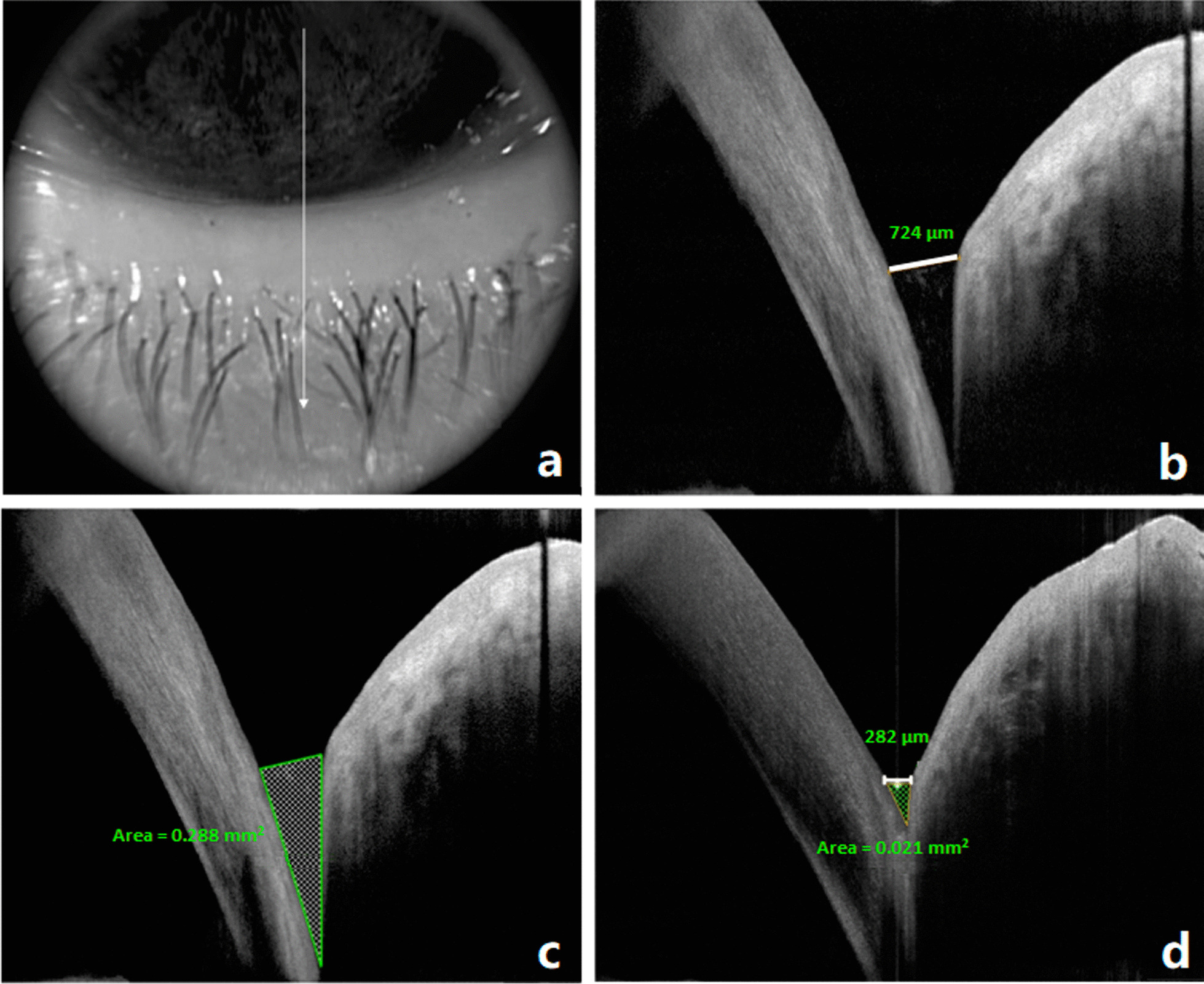
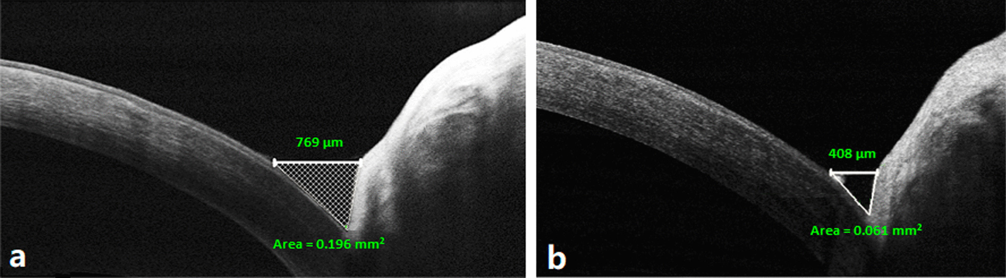
Methods: In a prospective study, the lower punctum of 54 eyes of 29 patients implanted with perforated punctal plugs were examined using AS-OCT during the early postoperative period. Preoperative tear meniscus height (TMH) and tear meniscus area (TMA) were evaluated. Postoperatively, the patency of the plug, its position, TMH and TMA were evaluated, and the results were correlated with postoperative epiphora. Munk scale was used for epiphora grading.
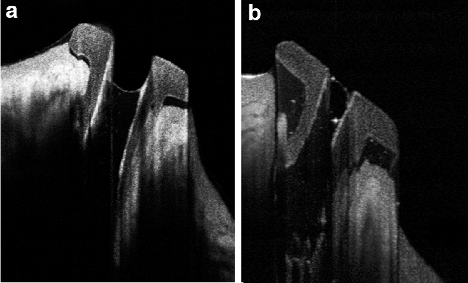
Results: Using AS-OCT, 48 (88.9%) plugs were found in proper position while 6 (11.1%) were rotated. The lumen of the plugs was completely patent in 47 (87%) plugs, partially obstructed in 2 (3.7%) plugs and completely occluded in 5 (9.2%) plugs. There was a statistically significant postoperative decrease of TMH and TMA (P < 0.001) and postoperative epiphora Munk score (P < 0.001).
Conclusion: AS-OCT is a valuable, reliable, and noninvasive investigative tool that can detect the proper positioning, patency, and contents of the implanted perforated lacrimal punctal plugs in addition to measurement of TMH and TMA. Trial registration ClinicalTrials.gov ID: NCT04624022, https://clinicaltrials.gov/ct2/show/NCT04624022.

 求助内容:
求助内容: 应助结果提醒方式:


