{"title":"乳腺血管肉瘤的造影诊断。","authors":"Maya Grisaru Kacen, Nikhil Sangle, Anat Kornecki","doi":"10.1155/2021/5542786","DOIUrl":null,"url":null,"abstract":"<p><p>A 60-year-old female presented for further assessment of a new right breast lump (November 2020). She had a history of a stage I (T1bN0M0) right breast invasive mammary carcinoma, grade 2 (score 7/9) with receptors ER/PR-negative, HER2/neu-positive, diagnosed four years prior to her current presentation. At that time, she was treated with a right breast lumpectomy and local radiation. Breast assessment with contrast-enhanced mammography showed new skin thickening with associated enhancement within the palpable region. Histology of subsequent ultrasound-guided biopsy found radiation-induced breast angiosarcoma. Breast angiosarcoma is a rare entity that represents less than 1% of all breast cancers. To our knowledge, this is the first case describing the imaging findings of breast angiosarcoma on contrast-enhanced mammography.</p>","PeriodicalId":30326,"journal":{"name":"Case Reports in Radiology","volume":" ","pages":"5542786"},"PeriodicalIF":0.0000,"publicationDate":"2021-08-12","publicationTypes":"Journal Article","fieldsOfStudy":null,"isOpenAccess":false,"openAccessPdf":"https://www.ncbi.nlm.nih.gov/pmc/articles/PMC8378973/pdf/","citationCount":"1","resultStr":"{\"title\":\"Contrast-Enhanced Mammography in the Diagnosis of Breast Angiosarcoma.\",\"authors\":\"Maya Grisaru Kacen, Nikhil Sangle, Anat Kornecki\",\"doi\":\"10.1155/2021/5542786\",\"DOIUrl\":null,\"url\":null,\"abstract\":\"<p><p>A 60-year-old female presented for further assessment of a new right breast lump (November 2020). She had a history of a stage I (T1bN0M0) right breast invasive mammary carcinoma, grade 2 (score 7/9) with receptors ER/PR-negative, HER2/neu-positive, diagnosed four years prior to her current presentation. At that time, she was treated with a right breast lumpectomy and local radiation. Breast assessment with contrast-enhanced mammography showed new skin thickening with associated enhancement within the palpable region. Histology of subsequent ultrasound-guided biopsy found radiation-induced breast angiosarcoma. Breast angiosarcoma is a rare entity that represents less than 1% of all breast cancers. To our knowledge, this is the first case describing the imaging findings of breast angiosarcoma on contrast-enhanced mammography.</p>\",\"PeriodicalId\":30326,\"journal\":{\"name\":\"Case Reports in Radiology\",\"volume\":\" \",\"pages\":\"5542786\"},\"PeriodicalIF\":0.0000,\"publicationDate\":\"2021-08-12\",\"publicationTypes\":\"Journal Article\",\"fieldsOfStudy\":null,\"isOpenAccess\":false,\"openAccessPdf\":\"https://www.ncbi.nlm.nih.gov/pmc/articles/PMC8378973/pdf/\",\"citationCount\":\"1\",\"resultStr\":null,\"platform\":\"Semanticscholar\",\"paperid\":null,\"PeriodicalName\":\"Case Reports in Radiology\",\"FirstCategoryId\":\"1085\",\"ListUrlMain\":\"https://doi.org/10.1155/2021/5542786\",\"RegionNum\":0,\"RegionCategory\":null,\"ArticlePicture\":[],\"TitleCN\":null,\"AbstractTextCN\":null,\"PMCID\":null,\"EPubDate\":\"2021/1/1 0:00:00\",\"PubModel\":\"eCollection\",\"JCR\":\"\",\"JCRName\":\"\",\"Score\":null,\"Total\":0}","platform":"Semanticscholar","paperid":null,"PeriodicalName":"Case Reports in Radiology","FirstCategoryId":"1085","ListUrlMain":"https://doi.org/10.1155/2021/5542786","RegionNum":0,"RegionCategory":null,"ArticlePicture":[],"TitleCN":null,"AbstractTextCN":null,"PMCID":null,"EPubDate":"2021/1/1 0:00:00","PubModel":"eCollection","JCR":"","JCRName":"","Score":null,"Total":0}
Contrast-Enhanced Mammography in the Diagnosis of Breast Angiosarcoma.
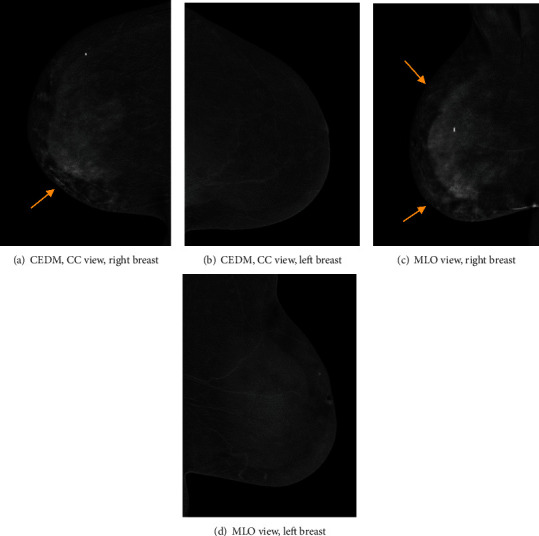
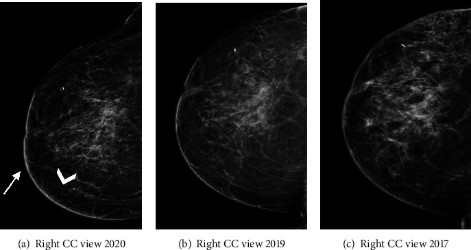
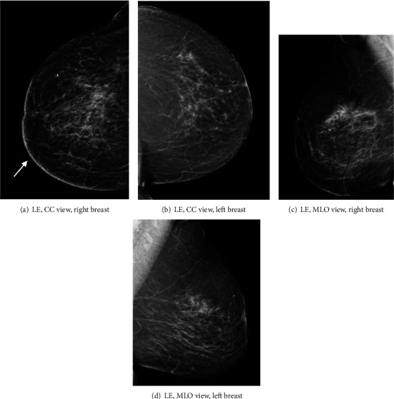
A 60-year-old female presented for further assessment of a new right breast lump (November 2020). She had a history of a stage I (T1bN0M0) right breast invasive mammary carcinoma, grade 2 (score 7/9) with receptors ER/PR-negative, HER2/neu-positive, diagnosed four years prior to her current presentation. At that time, she was treated with a right breast lumpectomy and local radiation. Breast assessment with contrast-enhanced mammography showed new skin thickening with associated enhancement within the palpable region. Histology of subsequent ultrasound-guided biopsy found radiation-induced breast angiosarcoma. Breast angiosarcoma is a rare entity that represents less than 1% of all breast cancers. To our knowledge, this is the first case describing the imaging findings of breast angiosarcoma on contrast-enhanced mammography.

 求助内容:
求助内容: 应助结果提醒方式:


