Josien Levenga, Helen Wong, Ryan Milstead, Lauren LaPlante, Charles A Hoeffer
{"title":"大脑中AKT亚型的免疫组织学检查:细胞类型特异性可能是AKT在复杂脑部疾病和神经系统疾病中作用的基础。","authors":"Josien Levenga, Helen Wong, Ryan Milstead, Lauren LaPlante, Charles A Hoeffer","doi":"10.1093/texcom/tgab036","DOIUrl":null,"url":null,"abstract":"<p><p>Protein kinase B (PKB/AKT) is a central kinase involved in many neurobiological processes. AKT is expressed in the brain as three isoforms, AKT1, AKT2, and AKT3. Previous studies suggest isoform-specific roles in neural function, but very few studies have examined AKT isoform expression at the cellular level. In this study, we use a combination of histology, immunostaining, and genetics to characterize cell-type-specific expression of AKT isoforms in human and mouse brains. In mice, we find that AKT1 is the most broadly expressed isoform, with expression in excitatory neurons and the sole detectable AKT isoform in gamma-aminobutyric acid ergic interneurons and microglia. By contrast, we find that AKT2 is the sole isoform expressed in astroglia and is not detected in other neural cell types. We find that AKT3 is expressed in excitatory neurons with AKT1 but shows greater expression levels in dendritic compartments than AKT1. We extend our analysis to human brain tissues and find similar results. Using genetic deletion approaches, we also find that the cellular determinants restricting AKT isoform expression to specific cell types remain intact under <i>Akt</i> deficiency conditions. Because AKT signaling is linked to numerous neurological disorders, a greater understanding of cell-specific isoform expression could improve treatment strategies involving AKT.</p>","PeriodicalId":72551,"journal":{"name":"Cerebral cortex communications","volume":" ","pages":"tgab036"},"PeriodicalIF":0.0000,"publicationDate":"2021-05-28","publicationTypes":"Journal Article","fieldsOfStudy":null,"isOpenAccess":false,"openAccessPdf":"https://www.ncbi.nlm.nih.gov/pmc/articles/PMC8223503/pdf/","citationCount":"8","resultStr":"{\"title\":\"Immunohistological Examination of AKT Isoforms in the Brain: Cell-Type Specificity That May Underlie AKT's Role in Complex Brain Disorders and Neurological Disease.\",\"authors\":\"Josien Levenga, Helen Wong, Ryan Milstead, Lauren LaPlante, Charles A Hoeffer\",\"doi\":\"10.1093/texcom/tgab036\",\"DOIUrl\":null,\"url\":null,\"abstract\":\"<p><p>Protein kinase B (PKB/AKT) is a central kinase involved in many neurobiological processes. AKT is expressed in the brain as three isoforms, AKT1, AKT2, and AKT3. Previous studies suggest isoform-specific roles in neural function, but very few studies have examined AKT isoform expression at the cellular level. In this study, we use a combination of histology, immunostaining, and genetics to characterize cell-type-specific expression of AKT isoforms in human and mouse brains. In mice, we find that AKT1 is the most broadly expressed isoform, with expression in excitatory neurons and the sole detectable AKT isoform in gamma-aminobutyric acid ergic interneurons and microglia. By contrast, we find that AKT2 is the sole isoform expressed in astroglia and is not detected in other neural cell types. We find that AKT3 is expressed in excitatory neurons with AKT1 but shows greater expression levels in dendritic compartments than AKT1. We extend our analysis to human brain tissues and find similar results. Using genetic deletion approaches, we also find that the cellular determinants restricting AKT isoform expression to specific cell types remain intact under <i>Akt</i> deficiency conditions. Because AKT signaling is linked to numerous neurological disorders, a greater understanding of cell-specific isoform expression could improve treatment strategies involving AKT.</p>\",\"PeriodicalId\":72551,\"journal\":{\"name\":\"Cerebral cortex communications\",\"volume\":\" \",\"pages\":\"tgab036\"},\"PeriodicalIF\":0.0000,\"publicationDate\":\"2021-05-28\",\"publicationTypes\":\"Journal Article\",\"fieldsOfStudy\":null,\"isOpenAccess\":false,\"openAccessPdf\":\"https://www.ncbi.nlm.nih.gov/pmc/articles/PMC8223503/pdf/\",\"citationCount\":\"8\",\"resultStr\":null,\"platform\":\"Semanticscholar\",\"paperid\":null,\"PeriodicalName\":\"Cerebral cortex communications\",\"FirstCategoryId\":\"1085\",\"ListUrlMain\":\"https://doi.org/10.1093/texcom/tgab036\",\"RegionNum\":0,\"RegionCategory\":null,\"ArticlePicture\":[],\"TitleCN\":null,\"AbstractTextCN\":null,\"PMCID\":null,\"EPubDate\":\"2021/1/1 0:00:00\",\"PubModel\":\"eCollection\",\"JCR\":\"\",\"JCRName\":\"\",\"Score\":null,\"Total\":0}","platform":"Semanticscholar","paperid":null,"PeriodicalName":"Cerebral cortex communications","FirstCategoryId":"1085","ListUrlMain":"https://doi.org/10.1093/texcom/tgab036","RegionNum":0,"RegionCategory":null,"ArticlePicture":[],"TitleCN":null,"AbstractTextCN":null,"PMCID":null,"EPubDate":"2021/1/1 0:00:00","PubModel":"eCollection","JCR":"","JCRName":"","Score":null,"Total":0}
Immunohistological Examination of AKT Isoforms in the Brain: Cell-Type Specificity That May Underlie AKT's Role in Complex Brain Disorders and Neurological Disease.
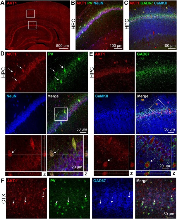
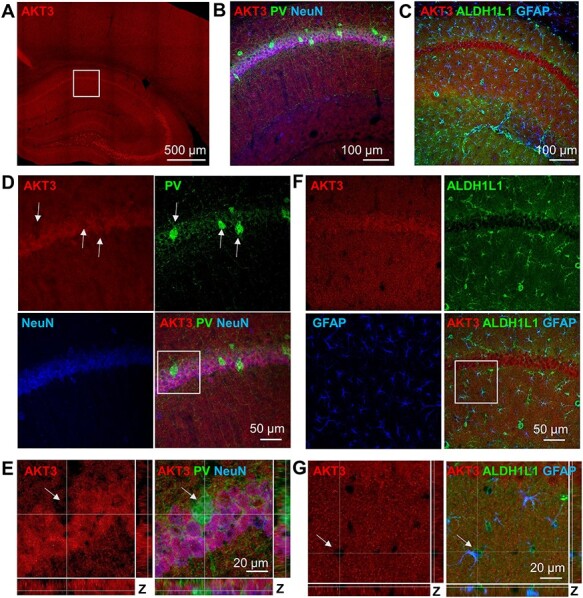
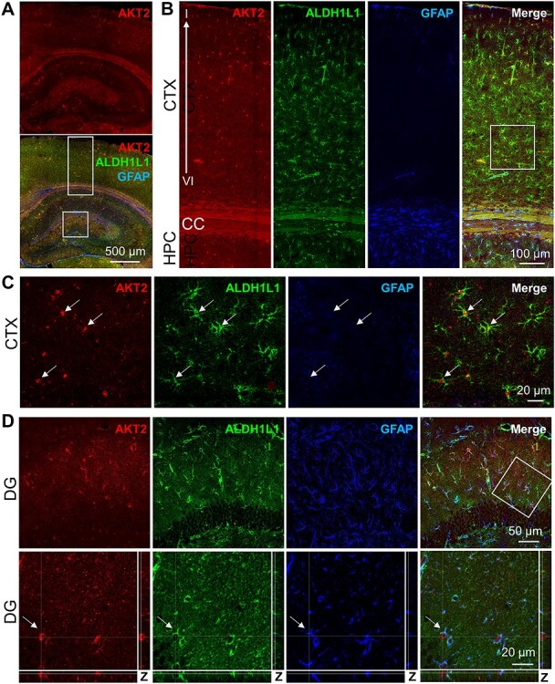
Protein kinase B (PKB/AKT) is a central kinase involved in many neurobiological processes. AKT is expressed in the brain as three isoforms, AKT1, AKT2, and AKT3. Previous studies suggest isoform-specific roles in neural function, but very few studies have examined AKT isoform expression at the cellular level. In this study, we use a combination of histology, immunostaining, and genetics to characterize cell-type-specific expression of AKT isoforms in human and mouse brains. In mice, we find that AKT1 is the most broadly expressed isoform, with expression in excitatory neurons and the sole detectable AKT isoform in gamma-aminobutyric acid ergic interneurons and microglia. By contrast, we find that AKT2 is the sole isoform expressed in astroglia and is not detected in other neural cell types. We find that AKT3 is expressed in excitatory neurons with AKT1 but shows greater expression levels in dendritic compartments than AKT1. We extend our analysis to human brain tissues and find similar results. Using genetic deletion approaches, we also find that the cellular determinants restricting AKT isoform expression to specific cell types remain intact under Akt deficiency conditions. Because AKT signaling is linked to numerous neurological disorders, a greater understanding of cell-specific isoform expression could improve treatment strategies involving AKT.

 求助内容:
求助内容: 应助结果提醒方式:


