快速扩张的主动脉根部真菌性假性动脉瘤伴流出道瘘。
引用次数: 0
摘要
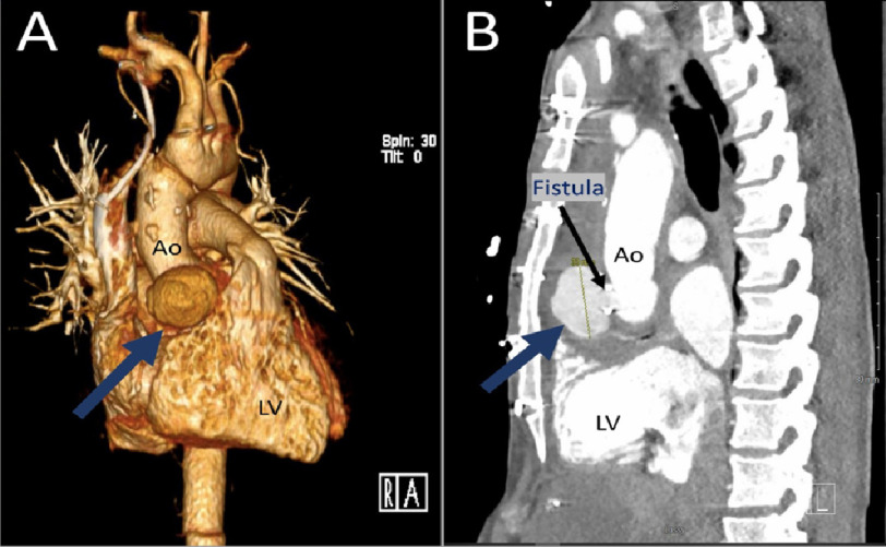
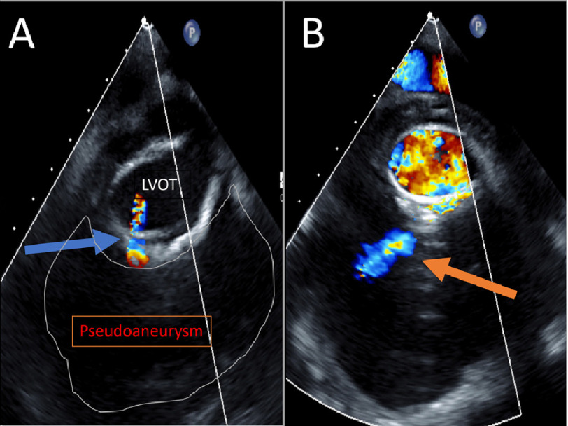
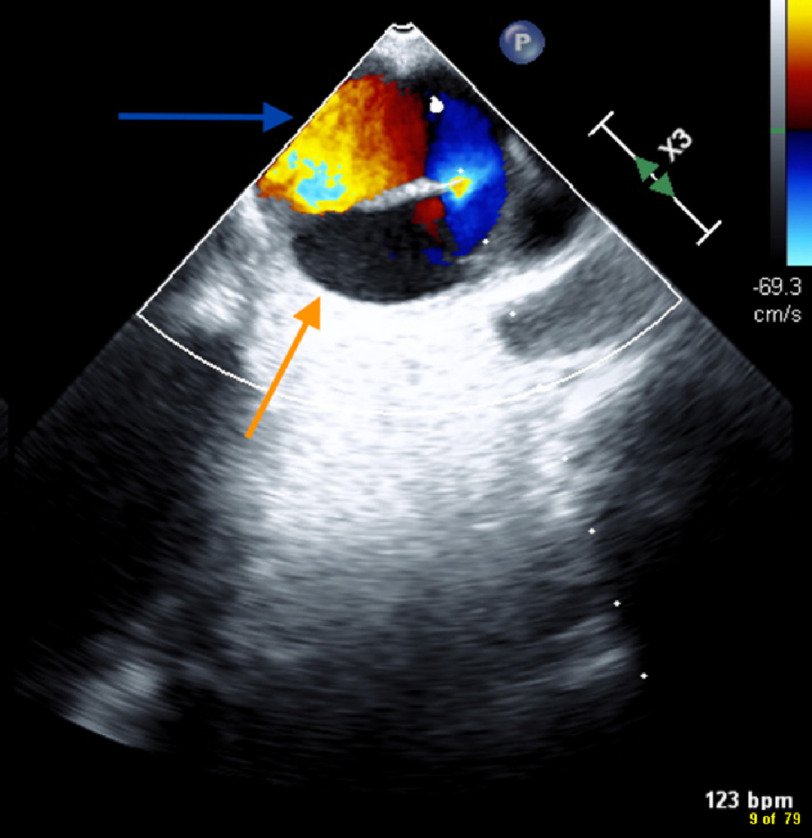
我们报告一位50岁的慢性Stanford a型主动脉夹层、感染性心内膜炎和快速扩张的主动脉周围肌细胞性假性动脉瘤伴LVOT瘘的患者。本病例强调了多模态成像在病理解剖复杂病例评估中的作用。本文章由计算机程序翻译,如有差异,请以英文原文为准。
Rapidly expanding aortic root mycotic pseudoaneurysm with outflow tract fistula.
We present a 50-year-old patient with chronic Stanford type-A aortic dissection, infective endocarditis, and rapidly expanding peri-aortic myocytic pseudoaneurysm with LVOT fistula. This case highlights the role of multimodality imaging in pathoanatomically complex-case evaluation.
求助全文
通过发布文献求助,成功后即可免费获取论文全文。
去求助
来源期刊

Global Cardiology Science & Practice
Medicine-Cardiology and Cardiovascular Medicine
CiteScore
1.60
自引率
0.00%
发文量
20
 求助内容:
求助内容: 应助结果提醒方式:


