Branko Stefanovic , Lela Stefanovic , Zarko Manojlovic
{"title":"细胞中I型前胶原生物合成成像显示高度组织化机体的生物发生;Collagenosomes","authors":"Branko Stefanovic , Lela Stefanovic , Zarko Manojlovic","doi":"10.1016/j.mbplus.2021.100076","DOIUrl":null,"url":null,"abstract":"<div><p>Mechanistic aspects of type I procollagen biosynthesis in cells are poorly understood. To provide more insight into this process we designed a system to directly image type I procollagen biogenesis by co-expression of fluorescently labeled full size procollagen α1(I) and one α2(I) polypeptides. High resolution images show that collagen α1(I) and α2(I) polypeptides are produced in coordination in discrete structures on the ER membrane, which we termed the collagenosomes. Collagenosomes are disk shaped bodies, 0.5–1 μM in diameter and 200–400 nm thick, in the core of which folding of procollagen takes place. Collagenosomes are intimately associated with the ER membrane and their formation requires intact translational machinery, suggesting that they are the sites of nascent procollagen biogenesis. Collagenosomes show little co-localization with the COPII transport vesicles, which export type I procollagen from the ER, suggesting that these two structures are distinct.</p><p>LARP6 is the protein which regulates translation of type I collagen mRNAs. The characteristic organization of collagenosomes depends on binding of LARP6 to collagen mRNAs. Without LARP6 regulation, collagenosomes are poorly organized and the folding of α1(I) and α2(I) polypeptides into procollagen in their cores is diminished. This indicates that formation of collagenosomes is dependent on regulated translation of collagen mRNAs. In live cells the size, number and shape of collagenosomes show little change within several hours, suggesting that they are stable structures of type I procollagen biogenesis. This is the first report of structural organization of type I collagen biogenesis in collagenosomes, while the fluorescent reporter system based on simultaneous imaging of both type I collagen polypeptides will enable the detailed elucidation of their structure and function.</p></div>","PeriodicalId":52317,"journal":{"name":"Matrix Biology Plus","volume":"12 ","pages":"Article 100076"},"PeriodicalIF":0.0000,"publicationDate":"2021-12-01","publicationTypes":"Journal Article","fieldsOfStudy":null,"isOpenAccess":false,"openAccessPdf":"https://sci-hub-pdf.com/10.1016/j.mbplus.2021.100076","citationCount":"2","resultStr":"{\"title\":\"Imaging of type I procollagen biosynthesis in cells reveals biogenesis in highly organized bodies; Collagenosomes\",\"authors\":\"Branko Stefanovic , Lela Stefanovic , Zarko Manojlovic\",\"doi\":\"10.1016/j.mbplus.2021.100076\",\"DOIUrl\":null,\"url\":null,\"abstract\":\"<div><p>Mechanistic aspects of type I procollagen biosynthesis in cells are poorly understood. To provide more insight into this process we designed a system to directly image type I procollagen biogenesis by co-expression of fluorescently labeled full size procollagen α1(I) and one α2(I) polypeptides. High resolution images show that collagen α1(I) and α2(I) polypeptides are produced in coordination in discrete structures on the ER membrane, which we termed the collagenosomes. Collagenosomes are disk shaped bodies, 0.5–1 μM in diameter and 200–400 nm thick, in the core of which folding of procollagen takes place. Collagenosomes are intimately associated with the ER membrane and their formation requires intact translational machinery, suggesting that they are the sites of nascent procollagen biogenesis. Collagenosomes show little co-localization with the COPII transport vesicles, which export type I procollagen from the ER, suggesting that these two structures are distinct.</p><p>LARP6 is the protein which regulates translation of type I collagen mRNAs. The characteristic organization of collagenosomes depends on binding of LARP6 to collagen mRNAs. Without LARP6 regulation, collagenosomes are poorly organized and the folding of α1(I) and α2(I) polypeptides into procollagen in their cores is diminished. This indicates that formation of collagenosomes is dependent on regulated translation of collagen mRNAs. In live cells the size, number and shape of collagenosomes show little change within several hours, suggesting that they are stable structures of type I procollagen biogenesis. This is the first report of structural organization of type I collagen biogenesis in collagenosomes, while the fluorescent reporter system based on simultaneous imaging of both type I collagen polypeptides will enable the detailed elucidation of their structure and function.</p></div>\",\"PeriodicalId\":52317,\"journal\":{\"name\":\"Matrix Biology Plus\",\"volume\":\"12 \",\"pages\":\"Article 100076\"},\"PeriodicalIF\":0.0000,\"publicationDate\":\"2021-12-01\",\"publicationTypes\":\"Journal Article\",\"fieldsOfStudy\":null,\"isOpenAccess\":false,\"openAccessPdf\":\"https://sci-hub-pdf.com/10.1016/j.mbplus.2021.100076\",\"citationCount\":\"2\",\"resultStr\":null,\"platform\":\"Semanticscholar\",\"paperid\":null,\"PeriodicalName\":\"Matrix Biology Plus\",\"FirstCategoryId\":\"1085\",\"ListUrlMain\":\"https://www.sciencedirect.com/science/article/pii/S259002852100020X\",\"RegionNum\":0,\"RegionCategory\":null,\"ArticlePicture\":[],\"TitleCN\":null,\"AbstractTextCN\":null,\"PMCID\":null,\"EPubDate\":\"\",\"PubModel\":\"\",\"JCR\":\"Q1\",\"JCRName\":\"Medicine\",\"Score\":null,\"Total\":0}","platform":"Semanticscholar","paperid":null,"PeriodicalName":"Matrix Biology Plus","FirstCategoryId":"1085","ListUrlMain":"https://www.sciencedirect.com/science/article/pii/S259002852100020X","RegionNum":0,"RegionCategory":null,"ArticlePicture":[],"TitleCN":null,"AbstractTextCN":null,"PMCID":null,"EPubDate":"","PubModel":"","JCR":"Q1","JCRName":"Medicine","Score":null,"Total":0}
Imaging of type I procollagen biosynthesis in cells reveals biogenesis in highly organized bodies; Collagenosomes
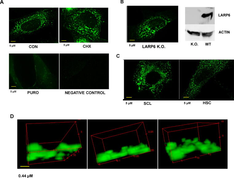
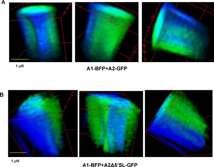
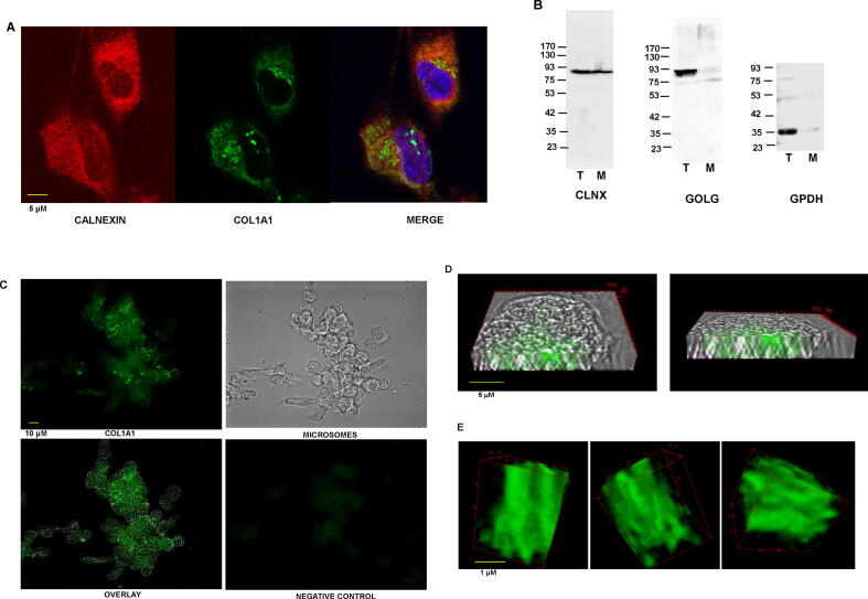
Mechanistic aspects of type I procollagen biosynthesis in cells are poorly understood. To provide more insight into this process we designed a system to directly image type I procollagen biogenesis by co-expression of fluorescently labeled full size procollagen α1(I) and one α2(I) polypeptides. High resolution images show that collagen α1(I) and α2(I) polypeptides are produced in coordination in discrete structures on the ER membrane, which we termed the collagenosomes. Collagenosomes are disk shaped bodies, 0.5–1 μM in diameter and 200–400 nm thick, in the core of which folding of procollagen takes place. Collagenosomes are intimately associated with the ER membrane and their formation requires intact translational machinery, suggesting that they are the sites of nascent procollagen biogenesis. Collagenosomes show little co-localization with the COPII transport vesicles, which export type I procollagen from the ER, suggesting that these two structures are distinct.
LARP6 is the protein which regulates translation of type I collagen mRNAs. The characteristic organization of collagenosomes depends on binding of LARP6 to collagen mRNAs. Without LARP6 regulation, collagenosomes are poorly organized and the folding of α1(I) and α2(I) polypeptides into procollagen in their cores is diminished. This indicates that formation of collagenosomes is dependent on regulated translation of collagen mRNAs. In live cells the size, number and shape of collagenosomes show little change within several hours, suggesting that they are stable structures of type I procollagen biogenesis. This is the first report of structural organization of type I collagen biogenesis in collagenosomes, while the fluorescent reporter system based on simultaneous imaging of both type I collagen polypeptides will enable the detailed elucidation of their structure and function.

 求助内容:
求助内容: 应助结果提醒方式:


