Francesco de Pasquale, Piero Chiacchiaretta, Luigi Pavone, Antonio Sparano, Paolo Capotosto, Giovanni Grillea, Giorgia Committeri, Antonello Baldassarre
{"title":"脑卒中后与视觉忽视相关的脑拓扑重组。","authors":"Francesco de Pasquale, Piero Chiacchiaretta, Luigi Pavone, Antonio Sparano, Paolo Capotosto, Giovanni Grillea, Giorgia Committeri, Antonello Baldassarre","doi":"10.1089/brain.2020.0969","DOIUrl":null,"url":null,"abstract":"<p><p><b><i>Background/Purpose:</i></b> To identify brain hubs that are behaviorally relevant for neglect after stroke as well as to characterize their functional architecture of communication. <b><i>Methods:</i></b> Twenty acute right hemisphere damaged patients underwent neuropsychological and resting-state functional magnetic resonance imaging sessions. Spatial neglect was assessed by means of the Center of Cancellation on the Bells Cancellation Test. For each patient, resting-state functional connectivity matrices were derived by adopting a brain parcellation scheme consisting of 153 nodes. For every node, we extracted its betweenness centrality (BC) defined as the portion of all shortest paths in the connectome involving such node. Then, neglect hubs were identified as those regions showing a high correlation between their BC and neglect scores. <b><i>Results:</i></b> A first set of neglect hubs was identified in multiple systems including dorsal attention and ventral attention, default mode, and frontoparietal executive-control networks within the damaged hemisphere as well as in the posterior and anterior cingulate cortex. Such cortical regions exhibited a loss of BC and increased (i.e., less efficient) weighted shortest path length (WSPL) related to severe neglect. Conversely, a second group of neglect hubs found in visual and motor networks, in the undamaged hemisphere, exhibited a pathological increase of BC and reduction of WSPL associated with severe neglect. <b><i>Conclusion:</i></b> The topological reorganization of the brain in neglect patients might reflect a maladaptive shift in processing spatial information from higher level associative-control systems to lower level visual and sensory-motor processing areas after a right hemisphere lesion.</p>","PeriodicalId":9155,"journal":{"name":"Brain connectivity","volume":" ","pages":"473-486"},"PeriodicalIF":2.4000,"publicationDate":"2023-10-01","publicationTypes":"Journal Article","fieldsOfStudy":null,"isOpenAccess":false,"openAccessPdf":"https://www.ncbi.nlm.nih.gov/pmc/articles/PMC10618825/pdf/","citationCount":"3","resultStr":"{\"title\":\"Brain Topological Reorganization Associated with Visual Neglect After Stroke.\",\"authors\":\"Francesco de Pasquale, Piero Chiacchiaretta, Luigi Pavone, Antonio Sparano, Paolo Capotosto, Giovanni Grillea, Giorgia Committeri, Antonello Baldassarre\",\"doi\":\"10.1089/brain.2020.0969\",\"DOIUrl\":null,\"url\":null,\"abstract\":\"<p><p><b><i>Background/Purpose:</i></b> To identify brain hubs that are behaviorally relevant for neglect after stroke as well as to characterize their functional architecture of communication. <b><i>Methods:</i></b> Twenty acute right hemisphere damaged patients underwent neuropsychological and resting-state functional magnetic resonance imaging sessions. Spatial neglect was assessed by means of the Center of Cancellation on the Bells Cancellation Test. For each patient, resting-state functional connectivity matrices were derived by adopting a brain parcellation scheme consisting of 153 nodes. For every node, we extracted its betweenness centrality (BC) defined as the portion of all shortest paths in the connectome involving such node. Then, neglect hubs were identified as those regions showing a high correlation between their BC and neglect scores. <b><i>Results:</i></b> A first set of neglect hubs was identified in multiple systems including dorsal attention and ventral attention, default mode, and frontoparietal executive-control networks within the damaged hemisphere as well as in the posterior and anterior cingulate cortex. Such cortical regions exhibited a loss of BC and increased (i.e., less efficient) weighted shortest path length (WSPL) related to severe neglect. Conversely, a second group of neglect hubs found in visual and motor networks, in the undamaged hemisphere, exhibited a pathological increase of BC and reduction of WSPL associated with severe neglect. <b><i>Conclusion:</i></b> The topological reorganization of the brain in neglect patients might reflect a maladaptive shift in processing spatial information from higher level associative-control systems to lower level visual and sensory-motor processing areas after a right hemisphere lesion.</p>\",\"PeriodicalId\":9155,\"journal\":{\"name\":\"Brain connectivity\",\"volume\":\" \",\"pages\":\"473-486\"},\"PeriodicalIF\":2.4000,\"publicationDate\":\"2023-10-01\",\"publicationTypes\":\"Journal Article\",\"fieldsOfStudy\":null,\"isOpenAccess\":false,\"openAccessPdf\":\"https://www.ncbi.nlm.nih.gov/pmc/articles/PMC10618825/pdf/\",\"citationCount\":\"3\",\"resultStr\":null,\"platform\":\"Semanticscholar\",\"paperid\":null,\"PeriodicalName\":\"Brain connectivity\",\"FirstCategoryId\":\"3\",\"ListUrlMain\":\"https://doi.org/10.1089/brain.2020.0969\",\"RegionNum\":3,\"RegionCategory\":\"医学\",\"ArticlePicture\":[],\"TitleCN\":null,\"AbstractTextCN\":null,\"PMCID\":null,\"EPubDate\":\"2021/9/15 0:00:00\",\"PubModel\":\"Epub\",\"JCR\":\"Q3\",\"JCRName\":\"NEUROSCIENCES\",\"Score\":null,\"Total\":0}","platform":"Semanticscholar","paperid":null,"PeriodicalName":"Brain connectivity","FirstCategoryId":"3","ListUrlMain":"https://doi.org/10.1089/brain.2020.0969","RegionNum":3,"RegionCategory":"医学","ArticlePicture":[],"TitleCN":null,"AbstractTextCN":null,"PMCID":null,"EPubDate":"2021/9/15 0:00:00","PubModel":"Epub","JCR":"Q3","JCRName":"NEUROSCIENCES","Score":null,"Total":0}
Brain Topological Reorganization Associated with Visual Neglect After Stroke.
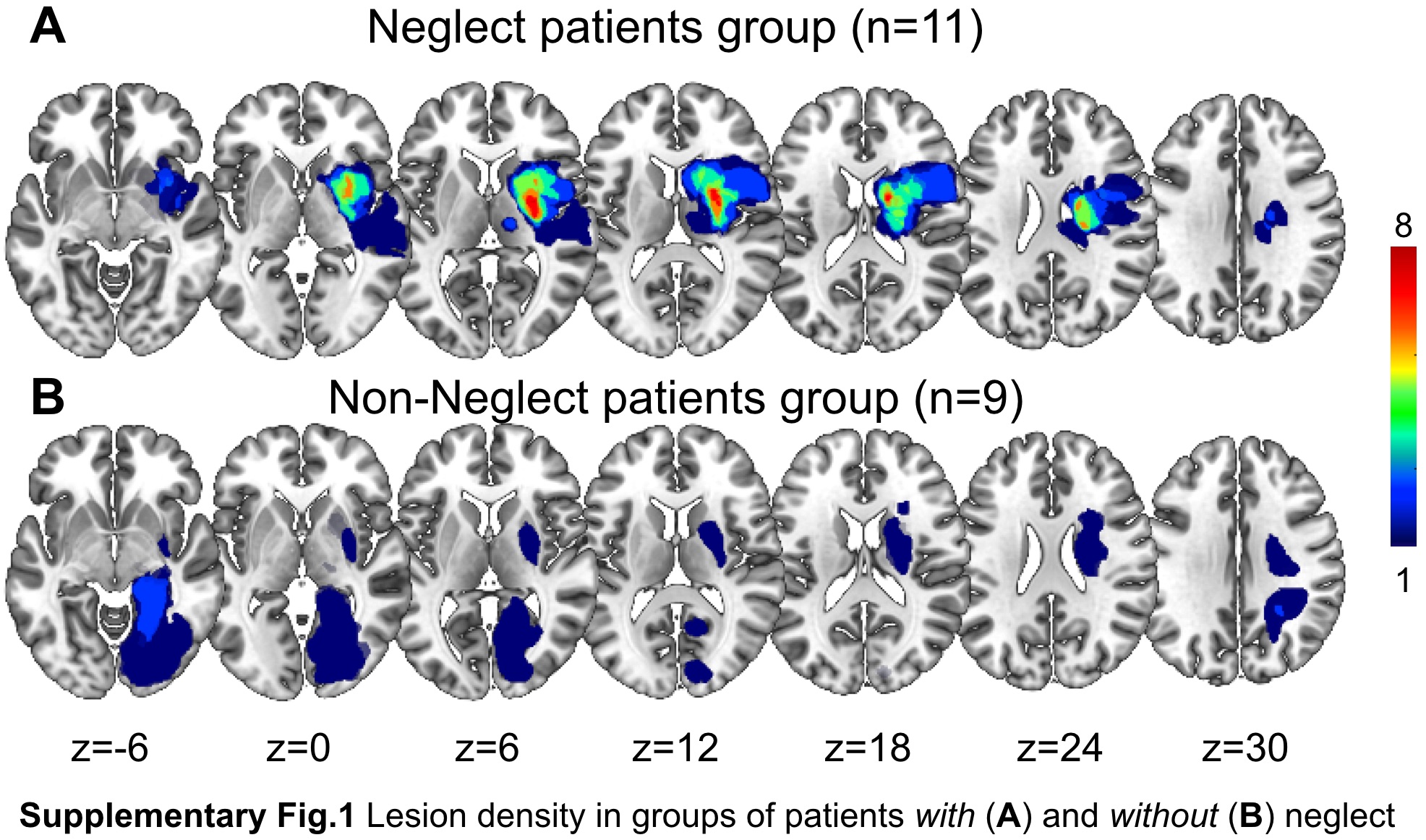
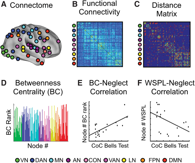
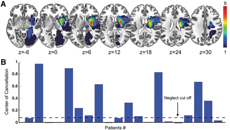
Background/Purpose: To identify brain hubs that are behaviorally relevant for neglect after stroke as well as to characterize their functional architecture of communication. Methods: Twenty acute right hemisphere damaged patients underwent neuropsychological and resting-state functional magnetic resonance imaging sessions. Spatial neglect was assessed by means of the Center of Cancellation on the Bells Cancellation Test. For each patient, resting-state functional connectivity matrices were derived by adopting a brain parcellation scheme consisting of 153 nodes. For every node, we extracted its betweenness centrality (BC) defined as the portion of all shortest paths in the connectome involving such node. Then, neglect hubs were identified as those regions showing a high correlation between their BC and neglect scores. Results: A first set of neglect hubs was identified in multiple systems including dorsal attention and ventral attention, default mode, and frontoparietal executive-control networks within the damaged hemisphere as well as in the posterior and anterior cingulate cortex. Such cortical regions exhibited a loss of BC and increased (i.e., less efficient) weighted shortest path length (WSPL) related to severe neglect. Conversely, a second group of neglect hubs found in visual and motor networks, in the undamaged hemisphere, exhibited a pathological increase of BC and reduction of WSPL associated with severe neglect. Conclusion: The topological reorganization of the brain in neglect patients might reflect a maladaptive shift in processing spatial information from higher level associative-control systems to lower level visual and sensory-motor processing areas after a right hemisphere lesion.
期刊介绍:
Brain Connectivity provides groundbreaking findings in the rapidly advancing field of connectivity research at the systems and network levels. The Journal disseminates information on brain mapping, modeling, novel research techniques, new imaging modalities, preclinical animal studies, and the translation of research discoveries from the laboratory to the clinic.
This essential journal fosters the application of basic biological discoveries and contributes to the development of novel diagnostic and therapeutic interventions to recognize and treat a broad range of neurodegenerative and psychiatric disorders such as: Alzheimer’s disease, attention-deficit hyperactivity disorder, posttraumatic stress disorder, epilepsy, traumatic brain injury, stroke, dementia, and depression.

 求助内容:
求助内容: 应助结果提醒方式:


