Lilian Aly, Eva-Maria Strauß, Nikolaus Feucht, Isabella Weiß, Achim Berthele, Meike Mitsdoerffer, Christian Haass, Bernhard Hemmer, Mathias Maier, Thomas Korn, Benjamin Knier
{"title":"光学相干断层扫描血管造影显示亚临床视网膜疾病的神经脊髓炎视谱障碍。","authors":"Lilian Aly, Eva-Maria Strauß, Nikolaus Feucht, Isabella Weiß, Achim Berthele, Meike Mitsdoerffer, Christian Haass, Bernhard Hemmer, Mathias Maier, Thomas Korn, Benjamin Knier","doi":"10.1177/13524585211028831","DOIUrl":null,"url":null,"abstract":"Background: Neuromyelitis optica spectrum disorders (NMOSD) are neuroinflammatory diseases of the central nervous system. Patients suffer from recurring relapses and it is unclear whether relapse-independent disease activity occurs and whether this is of clinical relevance. Objective: To detect disease-specific alterations of the retinal vasculature that reflect disease activity during NMOSD. Methods: Cross-sectional analysis of 16 patients with NMOSD, 21 patients with relapsing-remitting multiple sclerosis, and 21 healthy controls using retinal optical coherence tomography (OCT), optical coherence tomography angiography (OCT-A), measurement of glial fibrillary acidic protein (GFAP) serum levels, and assessment of visual acuity. Results: Patients with NMOSD but not multiple sclerosis revealed lower foveal thickness (FT) (p = 0.02) measures and an increase of the foveal avascular zone (FAZ) (p = 0.02) compared to healthy controls independent to optic neuritis. Reduced FT (p = 0.01), enlarged FAZ areas (p = 0.0001), and vessel loss of the superficial vascular complex (p = 0.01) were linked to higher serum GFAP levels and superficial vessel loss was associated with worse visual performance in patients with NMOSD irrespective of optic neuritis. Conclusion: Subclinical parafoveal retinal vessel loss might occur during NMOSD and might be linked to astrocyte damage and poor visual performance. OCT-A may be a tool to study subclinical disease activity during NMOSD.","PeriodicalId":520714,"journal":{"name":"Multiple sclerosis (Houndmills, Basingstoke, England)","volume":" ","pages":"522-531"},"PeriodicalIF":5.0000,"publicationDate":"2022-04-01","publicationTypes":"Journal Article","fieldsOfStudy":null,"isOpenAccess":false,"openAccessPdf":"https://www.ncbi.nlm.nih.gov/pmc/articles/PMC8961243/pdf/","citationCount":"18","resultStr":"{\"title\":\"Optical coherence tomography angiography indicates subclinical retinal disease in neuromyelitis optica spectrum disorders.\",\"authors\":\"Lilian Aly, Eva-Maria Strauß, Nikolaus Feucht, Isabella Weiß, Achim Berthele, Meike Mitsdoerffer, Christian Haass, Bernhard Hemmer, Mathias Maier, Thomas Korn, Benjamin Knier\",\"doi\":\"10.1177/13524585211028831\",\"DOIUrl\":null,\"url\":null,\"abstract\":\"Background: Neuromyelitis optica spectrum disorders (NMOSD) are neuroinflammatory diseases of the central nervous system. Patients suffer from recurring relapses and it is unclear whether relapse-independent disease activity occurs and whether this is of clinical relevance. Objective: To detect disease-specific alterations of the retinal vasculature that reflect disease activity during NMOSD. Methods: Cross-sectional analysis of 16 patients with NMOSD, 21 patients with relapsing-remitting multiple sclerosis, and 21 healthy controls using retinal optical coherence tomography (OCT), optical coherence tomography angiography (OCT-A), measurement of glial fibrillary acidic protein (GFAP) serum levels, and assessment of visual acuity. Results: Patients with NMOSD but not multiple sclerosis revealed lower foveal thickness (FT) (p = 0.02) measures and an increase of the foveal avascular zone (FAZ) (p = 0.02) compared to healthy controls independent to optic neuritis. Reduced FT (p = 0.01), enlarged FAZ areas (p = 0.0001), and vessel loss of the superficial vascular complex (p = 0.01) were linked to higher serum GFAP levels and superficial vessel loss was associated with worse visual performance in patients with NMOSD irrespective of optic neuritis. Conclusion: Subclinical parafoveal retinal vessel loss might occur during NMOSD and might be linked to astrocyte damage and poor visual performance. OCT-A may be a tool to study subclinical disease activity during NMOSD.\",\"PeriodicalId\":520714,\"journal\":{\"name\":\"Multiple sclerosis (Houndmills, Basingstoke, England)\",\"volume\":\" \",\"pages\":\"522-531\"},\"PeriodicalIF\":5.0000,\"publicationDate\":\"2022-04-01\",\"publicationTypes\":\"Journal Article\",\"fieldsOfStudy\":null,\"isOpenAccess\":false,\"openAccessPdf\":\"https://www.ncbi.nlm.nih.gov/pmc/articles/PMC8961243/pdf/\",\"citationCount\":\"18\",\"resultStr\":null,\"platform\":\"Semanticscholar\",\"paperid\":null,\"PeriodicalName\":\"Multiple sclerosis (Houndmills, Basingstoke, England)\",\"FirstCategoryId\":\"3\",\"ListUrlMain\":\"https://doi.org/10.1177/13524585211028831\",\"RegionNum\":0,\"RegionCategory\":null,\"ArticlePicture\":[],\"TitleCN\":null,\"AbstractTextCN\":null,\"PMCID\":null,\"EPubDate\":\"2021/7/14 0:00:00\",\"PubModel\":\"Epub\",\"JCR\":\"\",\"JCRName\":\"\",\"Score\":null,\"Total\":0}","platform":"Semanticscholar","paperid":null,"PeriodicalName":"Multiple sclerosis (Houndmills, Basingstoke, England)","FirstCategoryId":"3","ListUrlMain":"https://doi.org/10.1177/13524585211028831","RegionNum":0,"RegionCategory":null,"ArticlePicture":[],"TitleCN":null,"AbstractTextCN":null,"PMCID":null,"EPubDate":"2021/7/14 0:00:00","PubModel":"Epub","JCR":"","JCRName":"","Score":null,"Total":0}
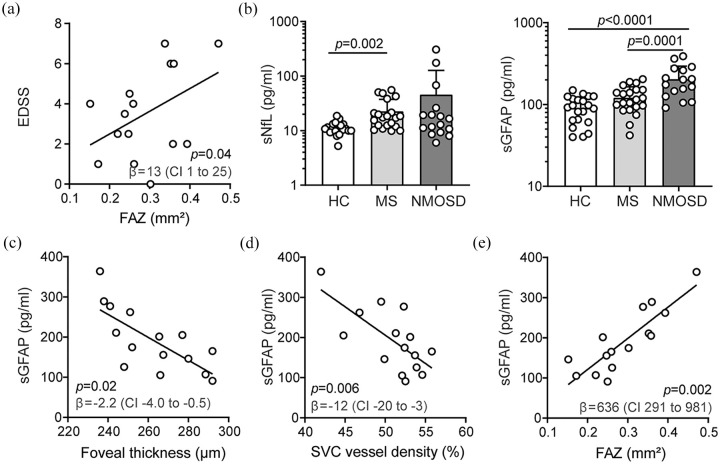
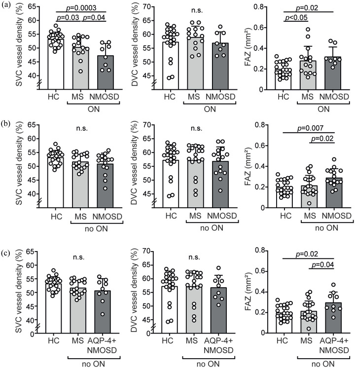
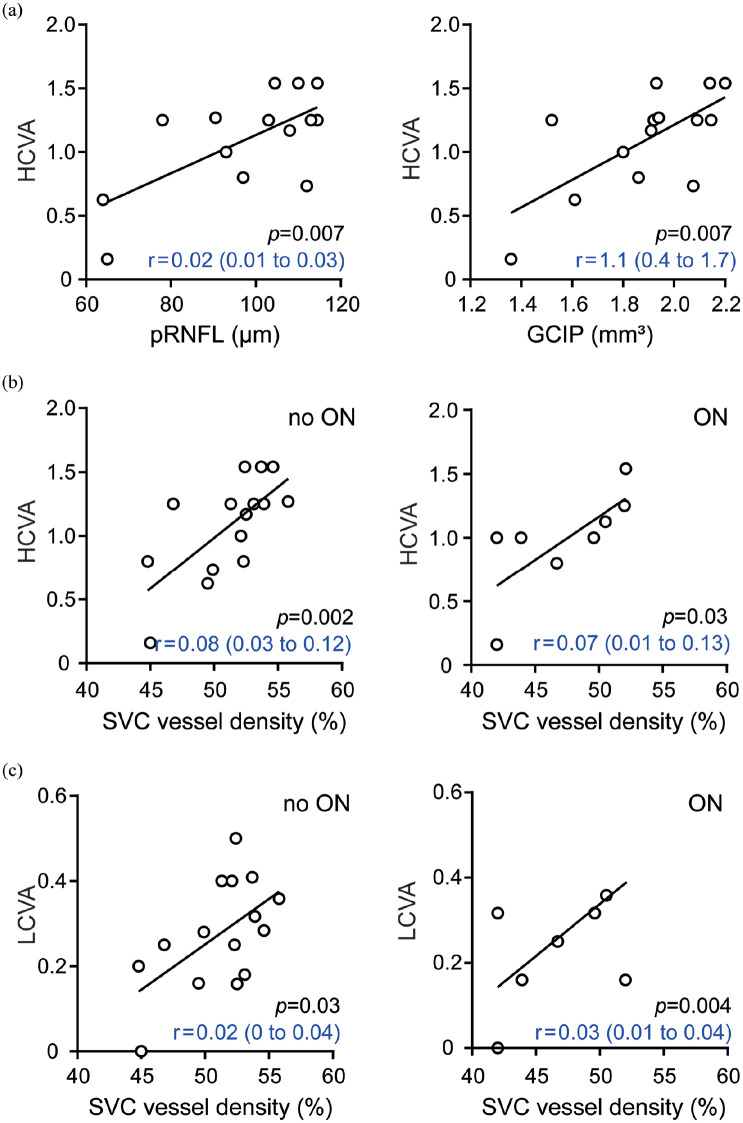
Background: Neuromyelitis optica spectrum disorders (NMOSD) are neuroinflammatory diseases of the central nervous system. Patients suffer from recurring relapses and it is unclear whether relapse-independent disease activity occurs and whether this is of clinical relevance. Objective: To detect disease-specific alterations of the retinal vasculature that reflect disease activity during NMOSD. Methods: Cross-sectional analysis of 16 patients with NMOSD, 21 patients with relapsing-remitting multiple sclerosis, and 21 healthy controls using retinal optical coherence tomography (OCT), optical coherence tomography angiography (OCT-A), measurement of glial fibrillary acidic protein (GFAP) serum levels, and assessment of visual acuity. Results: Patients with NMOSD but not multiple sclerosis revealed lower foveal thickness (FT) (p = 0.02) measures and an increase of the foveal avascular zone (FAZ) (p = 0.02) compared to healthy controls independent to optic neuritis. Reduced FT (p = 0.01), enlarged FAZ areas (p = 0.0001), and vessel loss of the superficial vascular complex (p = 0.01) were linked to higher serum GFAP levels and superficial vessel loss was associated with worse visual performance in patients with NMOSD irrespective of optic neuritis. Conclusion: Subclinical parafoveal retinal vessel loss might occur during NMOSD and might be linked to astrocyte damage and poor visual performance. OCT-A may be a tool to study subclinical disease activity during NMOSD.

 求助内容:
求助内容: 应助结果提醒方式:


