{"title":"颅内巨大脑膜瘤的外科治疗。","authors":"Soner Yaşar, Alparslan Kırık","doi":"10.5152/eurasianjmed.2021.20155","DOIUrl":null,"url":null,"abstract":"<p><strong>Objective: </strong>Giant intracranial meningiomas are a challenge for neurosurgeons because of their size and location in the cranium. Difficult tumor dissection and encasement of important neurovascular structures make them a horrible nightmare. The aims of this study are to present our giant intracranial meningioma series and to compare our experience using advanced surgical technology with the current literature.</p><p><strong>Materials and methods: </strong>The data of patients with the diagnosis of giant intracranial meningioma between 2014 and 2020 who underwent surgical treatment were retrospectively reviewed. The demographic, radiological, and surgical characteristics of patients were documented. The size and location of tumors as well as surgical technique were analyzed in detail.</p><p><strong>Results: </strong>A total of 61 patients with intracranial meningioma underwent surgical treatment over a 7-year period, and 10 (16.4%) tumors were larger than 5 cm in diameter, which were classified as giant meningioma. Seven patients were male and 3 were female, with a mean age of 64.9 years. Four tumors were located at the skull base. Histological diagnosis was meningioma World Health Organization grade I in 7 patients and grade II in 3 patients. Simpson grade 1 resection was achieved in 6 patients and grade 2 resection in 4 patients. No mortality was observed.</p><p><strong>Conclusion: </strong>Careful surgical planning should be made for giant intracranial meningiomas. Their location, adjacent neurovascular structures, and vascular supply affect the resection level of these giant tumors. Simpson grade 1 resection is seldom possible for skull base meningiomas.</p>","PeriodicalId":517142,"journal":{"name":"The Eurasian Journal of Medicine","volume":" ","pages":"73-78"},"PeriodicalIF":0.0000,"publicationDate":"2021-06-01","publicationTypes":"Journal Article","fieldsOfStudy":null,"isOpenAccess":false,"openAccessPdf":"https://www.ncbi.nlm.nih.gov/pmc/articles/PMC8184033/pdf/","citationCount":"3","resultStr":"{\"title\":\"Surgical Management of Giant Intracranial Meningiomas.\",\"authors\":\"Soner Yaşar, Alparslan Kırık\",\"doi\":\"10.5152/eurasianjmed.2021.20155\",\"DOIUrl\":null,\"url\":null,\"abstract\":\"<p><strong>Objective: </strong>Giant intracranial meningiomas are a challenge for neurosurgeons because of their size and location in the cranium. Difficult tumor dissection and encasement of important neurovascular structures make them a horrible nightmare. The aims of this study are to present our giant intracranial meningioma series and to compare our experience using advanced surgical technology with the current literature.</p><p><strong>Materials and methods: </strong>The data of patients with the diagnosis of giant intracranial meningioma between 2014 and 2020 who underwent surgical treatment were retrospectively reviewed. The demographic, radiological, and surgical characteristics of patients were documented. The size and location of tumors as well as surgical technique were analyzed in detail.</p><p><strong>Results: </strong>A total of 61 patients with intracranial meningioma underwent surgical treatment over a 7-year period, and 10 (16.4%) tumors were larger than 5 cm in diameter, which were classified as giant meningioma. Seven patients were male and 3 were female, with a mean age of 64.9 years. Four tumors were located at the skull base. Histological diagnosis was meningioma World Health Organization grade I in 7 patients and grade II in 3 patients. Simpson grade 1 resection was achieved in 6 patients and grade 2 resection in 4 patients. No mortality was observed.</p><p><strong>Conclusion: </strong>Careful surgical planning should be made for giant intracranial meningiomas. Their location, adjacent neurovascular structures, and vascular supply affect the resection level of these giant tumors. Simpson grade 1 resection is seldom possible for skull base meningiomas.</p>\",\"PeriodicalId\":517142,\"journal\":{\"name\":\"The Eurasian Journal of Medicine\",\"volume\":\" \",\"pages\":\"73-78\"},\"PeriodicalIF\":0.0000,\"publicationDate\":\"2021-06-01\",\"publicationTypes\":\"Journal Article\",\"fieldsOfStudy\":null,\"isOpenAccess\":false,\"openAccessPdf\":\"https://www.ncbi.nlm.nih.gov/pmc/articles/PMC8184033/pdf/\",\"citationCount\":\"3\",\"resultStr\":null,\"platform\":\"Semanticscholar\",\"paperid\":null,\"PeriodicalName\":\"The Eurasian Journal of Medicine\",\"FirstCategoryId\":\"1085\",\"ListUrlMain\":\"https://doi.org/10.5152/eurasianjmed.2021.20155\",\"RegionNum\":0,\"RegionCategory\":null,\"ArticlePicture\":[],\"TitleCN\":null,\"AbstractTextCN\":null,\"PMCID\":null,\"EPubDate\":\"\",\"PubModel\":\"\",\"JCR\":\"\",\"JCRName\":\"\",\"Score\":null,\"Total\":0}","platform":"Semanticscholar","paperid":null,"PeriodicalName":"The Eurasian Journal of Medicine","FirstCategoryId":"1085","ListUrlMain":"https://doi.org/10.5152/eurasianjmed.2021.20155","RegionNum":0,"RegionCategory":null,"ArticlePicture":[],"TitleCN":null,"AbstractTextCN":null,"PMCID":null,"EPubDate":"","PubModel":"","JCR":"","JCRName":"","Score":null,"Total":0}
Surgical Management of Giant Intracranial Meningiomas.
Objective: Giant intracranial meningiomas are a challenge for neurosurgeons because of their size and location in the cranium. Difficult tumor dissection and encasement of important neurovascular structures make them a horrible nightmare. The aims of this study are to present our giant intracranial meningioma series and to compare our experience using advanced surgical technology with the current literature.
Materials and methods: The data of patients with the diagnosis of giant intracranial meningioma between 2014 and 2020 who underwent surgical treatment were retrospectively reviewed. The demographic, radiological, and surgical characteristics of patients were documented. The size and location of tumors as well as surgical technique were analyzed in detail.
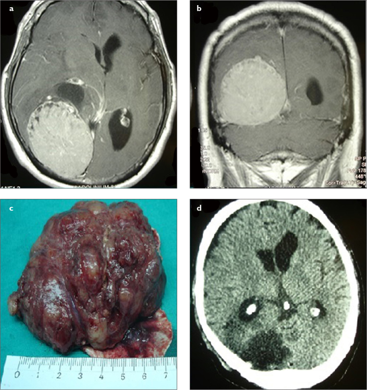
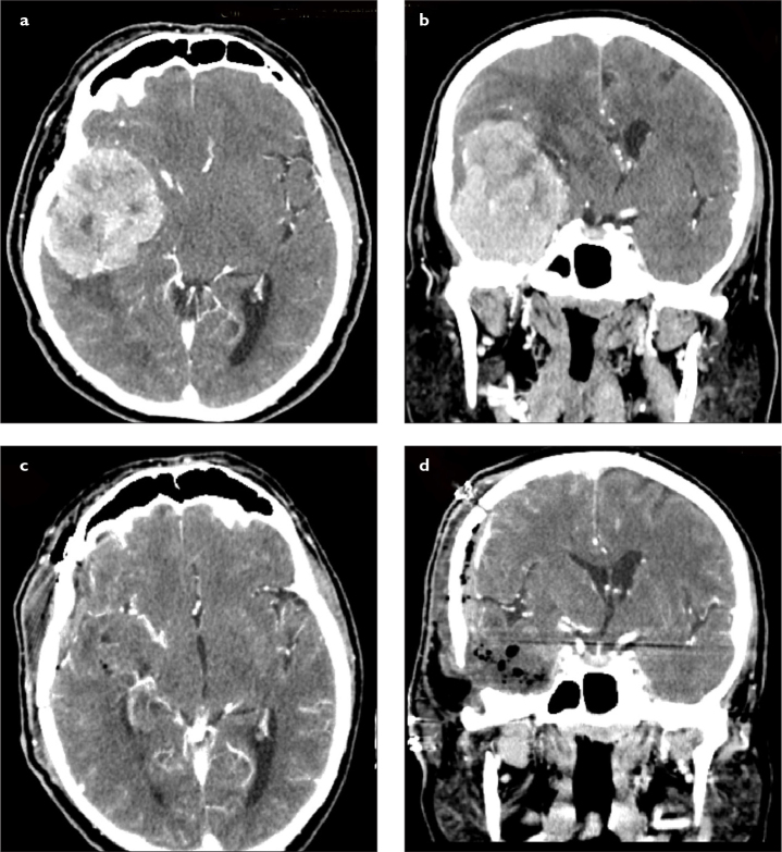
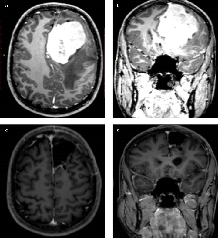
Results: A total of 61 patients with intracranial meningioma underwent surgical treatment over a 7-year period, and 10 (16.4%) tumors were larger than 5 cm in diameter, which were classified as giant meningioma. Seven patients were male and 3 were female, with a mean age of 64.9 years. Four tumors were located at the skull base. Histological diagnosis was meningioma World Health Organization grade I in 7 patients and grade II in 3 patients. Simpson grade 1 resection was achieved in 6 patients and grade 2 resection in 4 patients. No mortality was observed.
Conclusion: Careful surgical planning should be made for giant intracranial meningiomas. Their location, adjacent neurovascular structures, and vascular supply affect the resection level of these giant tumors. Simpson grade 1 resection is seldom possible for skull base meningiomas.

 求助内容:
求助内容: 应助结果提醒方式:


