Thomaz R Mostardeiro, Ananya Panda, Robert J Witte, Norbert G Campeau, Kiaran P McGee, Yi Sui, Aiming Lu
{"title":"全脑3D MR指纹脑成像:脑膜瘤患者的临床验证和可行性。","authors":"Thomaz R Mostardeiro, Ananya Panda, Robert J Witte, Norbert G Campeau, Kiaran P McGee, Yi Sui, Aiming Lu","doi":"10.1007/s10334-021-00924-1","DOIUrl":null,"url":null,"abstract":"<p><strong>Purpose: </strong>MR fingerprinting (MRF) is a MR technique that allows assessment of tissue relaxation times. The purpose of this study is to evaluate the clinical application of this technique in patients with meningioma.</p><p><strong>Materials and methods: </strong>A whole-brain 3D isotropic 1mm<sup>3</sup> acquisition under a 3.0T field strength was used to obtain MRF T<sub>1</sub> and T<sub>2</sub>-based relaxometry values in 4:38 s. The accuracy of values was quantified by scanning a quantitative MR relaxometry phantom. In vivo evaluation was performed by applying the sequence to 20 subjects with 25 meningiomas. Regions of interest included the meningioma, caudate head, centrum semiovale, contralateral white matter and thalamus. For both phantom and subjects, mean values of both T<sub>1</sub> and T<sub>2</sub> estimates were obtained. Statistical significance of differences in mean values between the meningioma and other brain structures was tested using a Friedman's ANOVA test.</p><p><strong>Results: </strong>MR fingerprinting phantom data demonstrated a linear relationship between measured and reference relaxometry estimates for both T<sub>1</sub> (r<sup>2</sup> = 0.99) and T<sub>2</sub> (r<sup>2</sup> = 0.97). MRF T<sub>1</sub> relaxation times were longer in meningioma (mean ± SD 1429 ± 202 ms) compared to thalamus (mean ± SD 1054 ± 58 ms; p = 0.004), centrum semiovale (mean ± SD 825 ± 42 ms; p < 0.001) and contralateral white matter (mean ± SD 799 ± 40 ms; p < 0.001). MRF T<sub>2</sub> relaxation times were longer for meningioma (mean ± SD 69 ± 27 ms) as compared to thalamus (mean ± SD 27 ± 3 ms; p < 0.001), caudate head (mean ± SD 39 ± 5 ms; p < 0.001) and contralateral white matter (mean ± SD 35 ± 4 ms; p < 0.001) CONCLUSIONS: Phantom measurements indicate that the proposed 3D-MRF sequence relaxometry estimations are valid and reproducible. For in vivo, entire brain coverage was obtained in clinically feasible time and allows quantitative assessment of meningioma in clinical practice.</p>","PeriodicalId":18067,"journal":{"name":"Magnetic Resonance Materials in Physics, Biology and Medicine","volume":"34 5","pages":"697-706"},"PeriodicalIF":2.0000,"publicationDate":"2021-10-01","publicationTypes":"Journal Article","fieldsOfStudy":null,"isOpenAccess":false,"openAccessPdf":"https://sci-hub-pdf.com/10.1007/s10334-021-00924-1","citationCount":"1","resultStr":"{\"title\":\"Whole-brain 3D MR fingerprinting brain imaging: clinical validation and feasibility to patients with meningioma.\",\"authors\":\"Thomaz R Mostardeiro, Ananya Panda, Robert J Witte, Norbert G Campeau, Kiaran P McGee, Yi Sui, Aiming Lu\",\"doi\":\"10.1007/s10334-021-00924-1\",\"DOIUrl\":null,\"url\":null,\"abstract\":\"<p><strong>Purpose: </strong>MR fingerprinting (MRF) is a MR technique that allows assessment of tissue relaxation times. The purpose of this study is to evaluate the clinical application of this technique in patients with meningioma.</p><p><strong>Materials and methods: </strong>A whole-brain 3D isotropic 1mm<sup>3</sup> acquisition under a 3.0T field strength was used to obtain MRF T<sub>1</sub> and T<sub>2</sub>-based relaxometry values in 4:38 s. The accuracy of values was quantified by scanning a quantitative MR relaxometry phantom. In vivo evaluation was performed by applying the sequence to 20 subjects with 25 meningiomas. Regions of interest included the meningioma, caudate head, centrum semiovale, contralateral white matter and thalamus. For both phantom and subjects, mean values of both T<sub>1</sub> and T<sub>2</sub> estimates were obtained. Statistical significance of differences in mean values between the meningioma and other brain structures was tested using a Friedman's ANOVA test.</p><p><strong>Results: </strong>MR fingerprinting phantom data demonstrated a linear relationship between measured and reference relaxometry estimates for both T<sub>1</sub> (r<sup>2</sup> = 0.99) and T<sub>2</sub> (r<sup>2</sup> = 0.97). MRF T<sub>1</sub> relaxation times were longer in meningioma (mean ± SD 1429 ± 202 ms) compared to thalamus (mean ± SD 1054 ± 58 ms; p = 0.004), centrum semiovale (mean ± SD 825 ± 42 ms; p < 0.001) and contralateral white matter (mean ± SD 799 ± 40 ms; p < 0.001). MRF T<sub>2</sub> relaxation times were longer for meningioma (mean ± SD 69 ± 27 ms) as compared to thalamus (mean ± SD 27 ± 3 ms; p < 0.001), caudate head (mean ± SD 39 ± 5 ms; p < 0.001) and contralateral white matter (mean ± SD 35 ± 4 ms; p < 0.001) CONCLUSIONS: Phantom measurements indicate that the proposed 3D-MRF sequence relaxometry estimations are valid and reproducible. For in vivo, entire brain coverage was obtained in clinically feasible time and allows quantitative assessment of meningioma in clinical practice.</p>\",\"PeriodicalId\":18067,\"journal\":{\"name\":\"Magnetic Resonance Materials in Physics, Biology and Medicine\",\"volume\":\"34 5\",\"pages\":\"697-706\"},\"PeriodicalIF\":2.0000,\"publicationDate\":\"2021-10-01\",\"publicationTypes\":\"Journal Article\",\"fieldsOfStudy\":null,\"isOpenAccess\":false,\"openAccessPdf\":\"https://sci-hub-pdf.com/10.1007/s10334-021-00924-1\",\"citationCount\":\"1\",\"resultStr\":null,\"platform\":\"Semanticscholar\",\"paperid\":null,\"PeriodicalName\":\"Magnetic Resonance Materials in Physics, Biology and Medicine\",\"FirstCategoryId\":\"3\",\"ListUrlMain\":\"https://doi.org/10.1007/s10334-021-00924-1\",\"RegionNum\":4,\"RegionCategory\":\"医学\",\"ArticlePicture\":[],\"TitleCN\":null,\"AbstractTextCN\":null,\"PMCID\":null,\"EPubDate\":\"2021/5/4 0:00:00\",\"PubModel\":\"Epub\",\"JCR\":\"Q3\",\"JCRName\":\"RADIOLOGY, NUCLEAR MEDICINE & MEDICAL IMAGING\",\"Score\":null,\"Total\":0}","platform":"Semanticscholar","paperid":null,"PeriodicalName":"Magnetic Resonance Materials in Physics, Biology and Medicine","FirstCategoryId":"3","ListUrlMain":"https://doi.org/10.1007/s10334-021-00924-1","RegionNum":4,"RegionCategory":"医学","ArticlePicture":[],"TitleCN":null,"AbstractTextCN":null,"PMCID":null,"EPubDate":"2021/5/4 0:00:00","PubModel":"Epub","JCR":"Q3","JCRName":"RADIOLOGY, NUCLEAR MEDICINE & MEDICAL IMAGING","Score":null,"Total":0}
Whole-brain 3D MR fingerprinting brain imaging: clinical validation and feasibility to patients with meningioma.
Purpose: MR fingerprinting (MRF) is a MR technique that allows assessment of tissue relaxation times. The purpose of this study is to evaluate the clinical application of this technique in patients with meningioma.
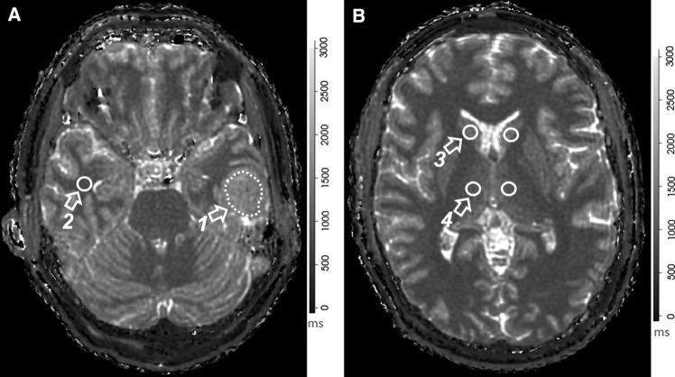
Materials and methods: A whole-brain 3D isotropic 1mm3 acquisition under a 3.0T field strength was used to obtain MRF T1 and T2-based relaxometry values in 4:38 s. The accuracy of values was quantified by scanning a quantitative MR relaxometry phantom. In vivo evaluation was performed by applying the sequence to 20 subjects with 25 meningiomas. Regions of interest included the meningioma, caudate head, centrum semiovale, contralateral white matter and thalamus. For both phantom and subjects, mean values of both T1 and T2 estimates were obtained. Statistical significance of differences in mean values between the meningioma and other brain structures was tested using a Friedman's ANOVA test.
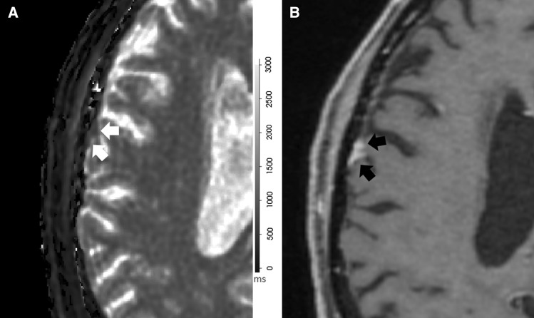
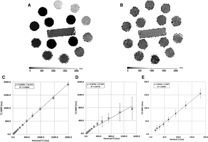
Results: MR fingerprinting phantom data demonstrated a linear relationship between measured and reference relaxometry estimates for both T1 (r2 = 0.99) and T2 (r2 = 0.97). MRF T1 relaxation times were longer in meningioma (mean ± SD 1429 ± 202 ms) compared to thalamus (mean ± SD 1054 ± 58 ms; p = 0.004), centrum semiovale (mean ± SD 825 ± 42 ms; p < 0.001) and contralateral white matter (mean ± SD 799 ± 40 ms; p < 0.001). MRF T2 relaxation times were longer for meningioma (mean ± SD 69 ± 27 ms) as compared to thalamus (mean ± SD 27 ± 3 ms; p < 0.001), caudate head (mean ± SD 39 ± 5 ms; p < 0.001) and contralateral white matter (mean ± SD 35 ± 4 ms; p < 0.001) CONCLUSIONS: Phantom measurements indicate that the proposed 3D-MRF sequence relaxometry estimations are valid and reproducible. For in vivo, entire brain coverage was obtained in clinically feasible time and allows quantitative assessment of meningioma in clinical practice.
期刊介绍:
MAGMA is a multidisciplinary international journal devoted to the publication of articles on all aspects of magnetic resonance techniques and their applications in medicine and biology. MAGMA currently publishes research papers, reviews, letters to the editor, and commentaries, six times a year. The subject areas covered by MAGMA include:
advances in materials, hardware and software in magnetic resonance technology,
new developments and results in research and practical applications of magnetic resonance imaging and spectroscopy related to biology and medicine,
study of animal models and intact cells using magnetic resonance,
reports of clinical trials on humans and clinical validation of magnetic resonance protocols.

 求助内容:
求助内容: 应助结果提醒方式:


