Céline Mahieu, Patrick Salvia, Benoît Beyer, Marcel Rooze, Véronique Feipel, Serge Van Sint Jan
{"title":"无症状受试者步态中跖弓变形和前足运动学。","authors":"Céline Mahieu, Patrick Salvia, Benoît Beyer, Marcel Rooze, Véronique Feipel, Serge Van Sint Jan","doi":"10.1080/23335432.2019.1642142","DOIUrl":null,"url":null,"abstract":"<p><p>This study aimed to investigate both foot arch-shaped architecture and forefoot kinematics during gait. Using a dedicated three-compartment forefoot subdivision, we studied asymptomatic subjects and quantified disorders related to the metatarsal arch. Foot motion and arch shape were measured in 30 healthy subjects with a motion-capture system and force plates. Kinematic results were expressed using a novel model, which anatomically divides the forefoot into three parts. This model integrated the medial longitudinal arch angle and the metatarsal arch height and width. During the first part of stance phase, the medial longitudinal arch flattens and all foot segments move toward dorsiflexion. During terminal stance and preswing phase, medial longitudinal and metatarsal arch restoration was noted with plantarflexion of all segments, an eversion and abduction of the medial forefoot, and an inversion and adduction of the lateral forefoot. Kinematics obtained with the proposed forefoot model corroborates metatarsal arch restoration in late stance. This observation supports the fact that foot architecture is supple until midstance and subsequently creates a rigid lever arm with restored arches to support propulsion. This study's results and methods highlight the potential of the three-compartment model for use in clinical decision-making.</p>","PeriodicalId":52124,"journal":{"name":"International Biomechanics","volume":"6 1","pages":"75-84"},"PeriodicalIF":0.0000,"publicationDate":"2019-12-01","publicationTypes":"Journal Article","fieldsOfStudy":null,"isOpenAccess":false,"openAccessPdf":"https://sci-hub-pdf.com/10.1080/23335432.2019.1642142","citationCount":"6","resultStr":"{\"title\":\"Metatarsal arch deformation and forefoot kinematics during gait in asymptomatic subjects.\",\"authors\":\"Céline Mahieu, Patrick Salvia, Benoît Beyer, Marcel Rooze, Véronique Feipel, Serge Van Sint Jan\",\"doi\":\"10.1080/23335432.2019.1642142\",\"DOIUrl\":null,\"url\":null,\"abstract\":\"<p><p>This study aimed to investigate both foot arch-shaped architecture and forefoot kinematics during gait. Using a dedicated three-compartment forefoot subdivision, we studied asymptomatic subjects and quantified disorders related to the metatarsal arch. Foot motion and arch shape were measured in 30 healthy subjects with a motion-capture system and force plates. Kinematic results were expressed using a novel model, which anatomically divides the forefoot into three parts. This model integrated the medial longitudinal arch angle and the metatarsal arch height and width. During the first part of stance phase, the medial longitudinal arch flattens and all foot segments move toward dorsiflexion. During terminal stance and preswing phase, medial longitudinal and metatarsal arch restoration was noted with plantarflexion of all segments, an eversion and abduction of the medial forefoot, and an inversion and adduction of the lateral forefoot. Kinematics obtained with the proposed forefoot model corroborates metatarsal arch restoration in late stance. This observation supports the fact that foot architecture is supple until midstance and subsequently creates a rigid lever arm with restored arches to support propulsion. This study's results and methods highlight the potential of the three-compartment model for use in clinical decision-making.</p>\",\"PeriodicalId\":52124,\"journal\":{\"name\":\"International Biomechanics\",\"volume\":\"6 1\",\"pages\":\"75-84\"},\"PeriodicalIF\":0.0000,\"publicationDate\":\"2019-12-01\",\"publicationTypes\":\"Journal Article\",\"fieldsOfStudy\":null,\"isOpenAccess\":false,\"openAccessPdf\":\"https://sci-hub-pdf.com/10.1080/23335432.2019.1642142\",\"citationCount\":\"6\",\"resultStr\":null,\"platform\":\"Semanticscholar\",\"paperid\":null,\"PeriodicalName\":\"International Biomechanics\",\"FirstCategoryId\":\"1085\",\"ListUrlMain\":\"https://doi.org/10.1080/23335432.2019.1642142\",\"RegionNum\":0,\"RegionCategory\":null,\"ArticlePicture\":[],\"TitleCN\":null,\"AbstractTextCN\":null,\"PMCID\":null,\"EPubDate\":\"\",\"PubModel\":\"\",\"JCR\":\"Q2\",\"JCRName\":\"Medicine\",\"Score\":null,\"Total\":0}","platform":"Semanticscholar","paperid":null,"PeriodicalName":"International Biomechanics","FirstCategoryId":"1085","ListUrlMain":"https://doi.org/10.1080/23335432.2019.1642142","RegionNum":0,"RegionCategory":null,"ArticlePicture":[],"TitleCN":null,"AbstractTextCN":null,"PMCID":null,"EPubDate":"","PubModel":"","JCR":"Q2","JCRName":"Medicine","Score":null,"Total":0}
Metatarsal arch deformation and forefoot kinematics during gait in asymptomatic subjects.
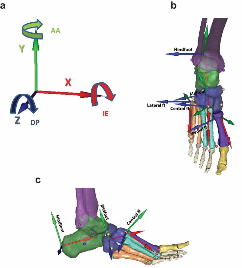
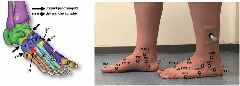
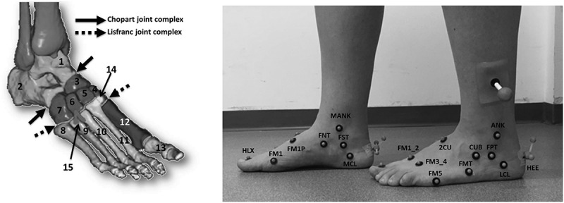
This study aimed to investigate both foot arch-shaped architecture and forefoot kinematics during gait. Using a dedicated three-compartment forefoot subdivision, we studied asymptomatic subjects and quantified disorders related to the metatarsal arch. Foot motion and arch shape were measured in 30 healthy subjects with a motion-capture system and force plates. Kinematic results were expressed using a novel model, which anatomically divides the forefoot into three parts. This model integrated the medial longitudinal arch angle and the metatarsal arch height and width. During the first part of stance phase, the medial longitudinal arch flattens and all foot segments move toward dorsiflexion. During terminal stance and preswing phase, medial longitudinal and metatarsal arch restoration was noted with plantarflexion of all segments, an eversion and abduction of the medial forefoot, and an inversion and adduction of the lateral forefoot. Kinematics obtained with the proposed forefoot model corroborates metatarsal arch restoration in late stance. This observation supports the fact that foot architecture is supple until midstance and subsequently creates a rigid lever arm with restored arches to support propulsion. This study's results and methods highlight the potential of the three-compartment model for use in clinical decision-making.
期刊介绍:
International Biomechanics is a fully Open Access biomechanics journal that aims to foster innovation, debate and collaboration across the full spectrum of biomechanics. We publish original articles, reviews, and short communications in all areas of biomechanics and welcome papers that explore: Bio-fluid mechanics, Continuum Biomechanics, Biotribology, Cellular Biomechanics, Mechanobiology, Mechano-transduction, Tissue Mechanics, Comparative Biomechanics and Functional Anatomy, Allometry, Animal locomotion in biomechanics, Gait analysis in biomechanics, Musculoskeletal and Orthopaedic Biomechanics, Cardiovascular Biomechanics, Plant Biomechanics, Injury Biomechanics, Impact Biomechanics, Sport and Exercise Biomechanics, Kinesiology, Rehabilitation in biomechanics, Quantitative Ergonomics, Human Factors engineering, Occupational Biomechanics, Developmental Biomechanics.
 求助内容:
求助内容: 应助结果提醒方式:


