Zeba Qadri, Valeria Righi, Shasha Li, A Aria Tzika
{"title":"利用分子磁共振成像在小鼠烧伤模型中追踪标记干细胞作为再生医学的一种方法。","authors":"Zeba Qadri, Valeria Righi, Shasha Li, A Aria Tzika","doi":"10.4236/ami.2021.111001","DOIUrl":null,"url":null,"abstract":"<p><p>Therapies based on stem cell transplants offer significant potential in the field of regenerative medicine. Monitoring the fate of the transplanted stem cells in a timely manner is considered one of the main limitations for long-standing success of stem cell transplants. Imaging methods that visualize and track stem cells <i>in vivo</i> non-invasively in real time are helpful towards the development of successful cell transplantation techniques. Novel molecular imaging methods which are non-invasive particularly such as MRI have been of great recent interest. Hence, mouse models which are of clinical relevance have been studied by injecting contrast agents used for labelling cells such as super-paramagnetic iron-oxide (SPIO) nanoparticles for cellular imaging. The MR techniques which can be used to generate positive contrast images have been of much relevance recently for tracking of the labelled cells. Particularly when the off-resonance region in the vicinity of the labeled cells is selectively excited while suppressing the signals from the non-labeled regions by the method of spectral dephasing. Thus, tracking of magnetically labelled cells employing positive contrast <i>in vivo</i> MR imaging methods in a burn mouse model in a non-invasive way has been the scope of this study. The consequences have direct implications for monitoring labeled stem cells at some stage in wound healing. We suggest that our approach can be used in clinical trials in molecular and regenerative medicine.</p>","PeriodicalId":64843,"journal":{"name":"分子影像学(英文)","volume":"11 1","pages":"1-15"},"PeriodicalIF":0.0000,"publicationDate":"2021-01-01","publicationTypes":"Journal Article","fieldsOfStudy":null,"isOpenAccess":false,"openAccessPdf":"https://www.ncbi.nlm.nih.gov/pmc/articles/PMC8118598/pdf/","citationCount":"2","resultStr":"{\"title\":\"Tracking of Labelled Stem Cells Using Molecular MR Imaging in a Mouse Burn Model <i>in Vivo</i> as an Approach to Regenerative Medicine.\",\"authors\":\"Zeba Qadri, Valeria Righi, Shasha Li, A Aria Tzika\",\"doi\":\"10.4236/ami.2021.111001\",\"DOIUrl\":null,\"url\":null,\"abstract\":\"<p><p>Therapies based on stem cell transplants offer significant potential in the field of regenerative medicine. Monitoring the fate of the transplanted stem cells in a timely manner is considered one of the main limitations for long-standing success of stem cell transplants. Imaging methods that visualize and track stem cells <i>in vivo</i> non-invasively in real time are helpful towards the development of successful cell transplantation techniques. Novel molecular imaging methods which are non-invasive particularly such as MRI have been of great recent interest. Hence, mouse models which are of clinical relevance have been studied by injecting contrast agents used for labelling cells such as super-paramagnetic iron-oxide (SPIO) nanoparticles for cellular imaging. The MR techniques which can be used to generate positive contrast images have been of much relevance recently for tracking of the labelled cells. Particularly when the off-resonance region in the vicinity of the labeled cells is selectively excited while suppressing the signals from the non-labeled regions by the method of spectral dephasing. Thus, tracking of magnetically labelled cells employing positive contrast <i>in vivo</i> MR imaging methods in a burn mouse model in a non-invasive way has been the scope of this study. The consequences have direct implications for monitoring labeled stem cells at some stage in wound healing. We suggest that our approach can be used in clinical trials in molecular and regenerative medicine.</p>\",\"PeriodicalId\":64843,\"journal\":{\"name\":\"分子影像学(英文)\",\"volume\":\"11 1\",\"pages\":\"1-15\"},\"PeriodicalIF\":0.0000,\"publicationDate\":\"2021-01-01\",\"publicationTypes\":\"Journal Article\",\"fieldsOfStudy\":null,\"isOpenAccess\":false,\"openAccessPdf\":\"https://www.ncbi.nlm.nih.gov/pmc/articles/PMC8118598/pdf/\",\"citationCount\":\"2\",\"resultStr\":null,\"platform\":\"Semanticscholar\",\"paperid\":null,\"PeriodicalName\":\"分子影像学(英文)\",\"FirstCategoryId\":\"1085\",\"ListUrlMain\":\"https://doi.org/10.4236/ami.2021.111001\",\"RegionNum\":0,\"RegionCategory\":null,\"ArticlePicture\":[],\"TitleCN\":null,\"AbstractTextCN\":null,\"PMCID\":null,\"EPubDate\":\"\",\"PubModel\":\"\",\"JCR\":\"\",\"JCRName\":\"\",\"Score\":null,\"Total\":0}","platform":"Semanticscholar","paperid":null,"PeriodicalName":"分子影像学(英文)","FirstCategoryId":"1085","ListUrlMain":"https://doi.org/10.4236/ami.2021.111001","RegionNum":0,"RegionCategory":null,"ArticlePicture":[],"TitleCN":null,"AbstractTextCN":null,"PMCID":null,"EPubDate":"","PubModel":"","JCR":"","JCRName":"","Score":null,"Total":0}
Tracking of Labelled Stem Cells Using Molecular MR Imaging in a Mouse Burn Model in Vivo as an Approach to Regenerative Medicine.
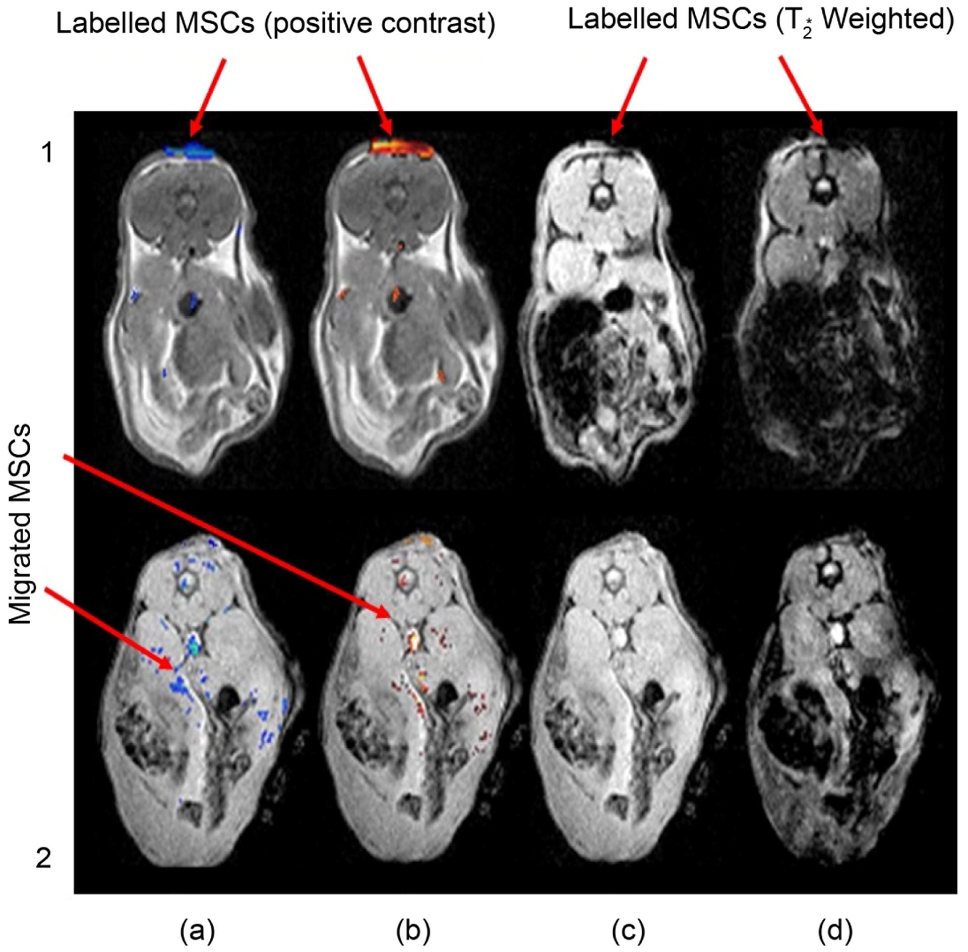
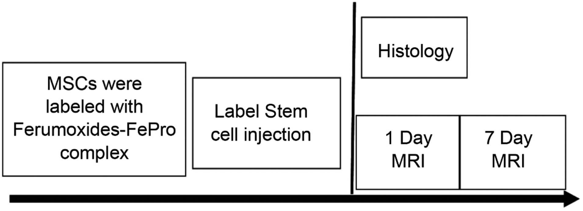
Therapies based on stem cell transplants offer significant potential in the field of regenerative medicine. Monitoring the fate of the transplanted stem cells in a timely manner is considered one of the main limitations for long-standing success of stem cell transplants. Imaging methods that visualize and track stem cells in vivo non-invasively in real time are helpful towards the development of successful cell transplantation techniques. Novel molecular imaging methods which are non-invasive particularly such as MRI have been of great recent interest. Hence, mouse models which are of clinical relevance have been studied by injecting contrast agents used for labelling cells such as super-paramagnetic iron-oxide (SPIO) nanoparticles for cellular imaging. The MR techniques which can be used to generate positive contrast images have been of much relevance recently for tracking of the labelled cells. Particularly when the off-resonance region in the vicinity of the labeled cells is selectively excited while suppressing the signals from the non-labeled regions by the method of spectral dephasing. Thus, tracking of magnetically labelled cells employing positive contrast in vivo MR imaging methods in a burn mouse model in a non-invasive way has been the scope of this study. The consequences have direct implications for monitoring labeled stem cells at some stage in wound healing. We suggest that our approach can be used in clinical trials in molecular and regenerative medicine.

 求助内容:
求助内容: 应助结果提醒方式:


