Alex Vicino, Valentin Loser, Paolo Salvioni Chiabotti, Jean Philippe Brouland, Renaud Du Pasquier
{"title":"抗腺苷酸激酶5脑炎与中枢神经系统血管炎的组织学证据。","authors":"Alex Vicino, Valentin Loser, Paolo Salvioni Chiabotti, Jean Philippe Brouland, Renaud Du Pasquier","doi":"10.1212/NXI.0000000000001010","DOIUrl":null,"url":null,"abstract":"Basic laboratory tests were normal. A brain MRI at admission (1 month after symptoms onset) showed bilateral, right predominant, mesiotemporal T2—fluid-attenuated inversion recovery (FLAIR) hyperintensity with gadolinium enhancement (figure). CSF analysis showed lymphocytic pleocytosis (120 cells/mm, 98% lymphocytes), hyperproteinorachia (1,032 mg/L), intrathecal IgG synthesis, and normal glucose and lactate levels. PCR for encephalitis, including HSV-1, was negative. Tuberculosis and Whipple disease were ruled out. Immunologic studies, CSF cytology, and flow cytometry were unremarkable.","PeriodicalId":520720,"journal":{"name":"Neurology(R) neuroimmunology & neuroinflammation","volume":" ","pages":""},"PeriodicalIF":7.5000,"publicationDate":"2021-05-11","publicationTypes":"Journal Article","fieldsOfStudy":null,"isOpenAccess":false,"openAccessPdf":"https://ftp.ncbi.nlm.nih.gov/pub/pmc/oa_pdf/02/9b/NEURIMMINFL2020038569.PMC8114832.pdf","citationCount":"2","resultStr":"{\"title\":\"Anti-Adenylate Kinase 5 Encephalitis With Histologic Evidence of CNS Vasculitis.\",\"authors\":\"Alex Vicino, Valentin Loser, Paolo Salvioni Chiabotti, Jean Philippe Brouland, Renaud Du Pasquier\",\"doi\":\"10.1212/NXI.0000000000001010\",\"DOIUrl\":null,\"url\":null,\"abstract\":\"Basic laboratory tests were normal. A brain MRI at admission (1 month after symptoms onset) showed bilateral, right predominant, mesiotemporal T2—fluid-attenuated inversion recovery (FLAIR) hyperintensity with gadolinium enhancement (figure). CSF analysis showed lymphocytic pleocytosis (120 cells/mm, 98% lymphocytes), hyperproteinorachia (1,032 mg/L), intrathecal IgG synthesis, and normal glucose and lactate levels. PCR for encephalitis, including HSV-1, was negative. Tuberculosis and Whipple disease were ruled out. Immunologic studies, CSF cytology, and flow cytometry were unremarkable.\",\"PeriodicalId\":520720,\"journal\":{\"name\":\"Neurology(R) neuroimmunology & neuroinflammation\",\"volume\":\" \",\"pages\":\"\"},\"PeriodicalIF\":7.5000,\"publicationDate\":\"2021-05-11\",\"publicationTypes\":\"Journal Article\",\"fieldsOfStudy\":null,\"isOpenAccess\":false,\"openAccessPdf\":\"https://ftp.ncbi.nlm.nih.gov/pub/pmc/oa_pdf/02/9b/NEURIMMINFL2020038569.PMC8114832.pdf\",\"citationCount\":\"2\",\"resultStr\":null,\"platform\":\"Semanticscholar\",\"paperid\":null,\"PeriodicalName\":\"Neurology(R) neuroimmunology & neuroinflammation\",\"FirstCategoryId\":\"3\",\"ListUrlMain\":\"https://doi.org/10.1212/NXI.0000000000001010\",\"RegionNum\":0,\"RegionCategory\":null,\"ArticlePicture\":[],\"TitleCN\":null,\"AbstractTextCN\":null,\"PMCID\":null,\"EPubDate\":\"2021/7/1 0:00:00\",\"PubModel\":\"Print\",\"JCR\":\"\",\"JCRName\":\"\",\"Score\":null,\"Total\":0}","platform":"Semanticscholar","paperid":null,"PeriodicalName":"Neurology(R) neuroimmunology & neuroinflammation","FirstCategoryId":"3","ListUrlMain":"https://doi.org/10.1212/NXI.0000000000001010","RegionNum":0,"RegionCategory":null,"ArticlePicture":[],"TitleCN":null,"AbstractTextCN":null,"PMCID":null,"EPubDate":"2021/7/1 0:00:00","PubModel":"Print","JCR":"","JCRName":"","Score":null,"Total":0}
Anti-Adenylate Kinase 5 Encephalitis With Histologic Evidence of CNS Vasculitis.
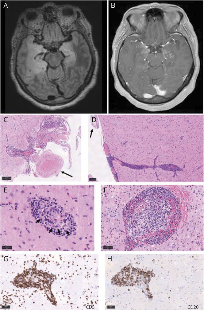
Basic laboratory tests were normal. A brain MRI at admission (1 month after symptoms onset) showed bilateral, right predominant, mesiotemporal T2—fluid-attenuated inversion recovery (FLAIR) hyperintensity with gadolinium enhancement (figure). CSF analysis showed lymphocytic pleocytosis (120 cells/mm, 98% lymphocytes), hyperproteinorachia (1,032 mg/L), intrathecal IgG synthesis, and normal glucose and lactate levels. PCR for encephalitis, including HSV-1, was negative. Tuberculosis and Whipple disease were ruled out. Immunologic studies, CSF cytology, and flow cytometry were unremarkable.

 求助内容:
求助内容: 应助结果提醒方式:


