Andresa Borges Soares, Vera Cavalcanti de Araújo, Fabricio Passador-Santos, Luiz Alexandre Thomaz, Andre Luis Santana de Freitas, Mario Claudio Mautoni, Rafael Fantelli Stelini, Maria Leticia Cintra
{"title":"罕见色素原位癌1例报告及简要回顾。","authors":"Andresa Borges Soares, Vera Cavalcanti de Araújo, Fabricio Passador-Santos, Luiz Alexandre Thomaz, Andre Luis Santana de Freitas, Mario Claudio Mautoni, Rafael Fantelli Stelini, Maria Leticia Cintra","doi":"10.1177/2632010X211009819","DOIUrl":null,"url":null,"abstract":"<p><p>Pigmented lesions of the oral mucosa encompass several benign and malignant conditions that may be a matter of concern under both clinical and histopathological views. We reported a case of a 62-year-old woman, presenting with an asymptomatic, deeply pigmented lesion on the soft palate. On examination, it appeared asymmetrical, with irregular borders and an area of ulceration. A biopsy, taken to rule out melanoma, revealed a pigmented carcinoma in situ. Throughout the tumor thickness, numerous interspersed melanocytes were found that did not extend to neighboring epithelium. These were large, richly dendritic, and presented abundance of melanin granules and small nuclei. Mild melanin incontinence was found. Scanty transfer of pigment to dysplastic epithelial cells was found through Fontana Masson staining. On immunohistochemical analyses, there were pancytokeratin-stained tumor epithelial cells; increased cell proliferation throughout the entire thickness of the tumor was emphasized by Ki-67 immunomarking. P16 was negative. The dendritic cells were selectively stained for S-100, HMB45 and Melan A. Wide spectrum in situ hybridization for human papillomavirus (HPV) was negative. Unfortunately, following diagnosis, the patient refused any treatment option. Pigmented squamous cell carcinoma with melanocyte colonization must be taken into account in the differential diagnosis of pigmented lesions of the oral cavity.</p>","PeriodicalId":53204,"journal":{"name":"Clinical Pathology","volume":"14 ","pages":"2632010X211009819"},"PeriodicalIF":1.9000,"publicationDate":"2021-04-20","publicationTypes":"Journal Article","fieldsOfStudy":null,"isOpenAccess":false,"openAccessPdf":"https://sci-hub-pdf.com/10.1177/2632010X211009819","citationCount":"0","resultStr":"{\"title\":\"Uncommon Pigmented Carcinoma In Situ: Case Report and Brief Review.\",\"authors\":\"Andresa Borges Soares, Vera Cavalcanti de Araújo, Fabricio Passador-Santos, Luiz Alexandre Thomaz, Andre Luis Santana de Freitas, Mario Claudio Mautoni, Rafael Fantelli Stelini, Maria Leticia Cintra\",\"doi\":\"10.1177/2632010X211009819\",\"DOIUrl\":null,\"url\":null,\"abstract\":\"<p><p>Pigmented lesions of the oral mucosa encompass several benign and malignant conditions that may be a matter of concern under both clinical and histopathological views. We reported a case of a 62-year-old woman, presenting with an asymptomatic, deeply pigmented lesion on the soft palate. On examination, it appeared asymmetrical, with irregular borders and an area of ulceration. A biopsy, taken to rule out melanoma, revealed a pigmented carcinoma in situ. Throughout the tumor thickness, numerous interspersed melanocytes were found that did not extend to neighboring epithelium. These were large, richly dendritic, and presented abundance of melanin granules and small nuclei. Mild melanin incontinence was found. Scanty transfer of pigment to dysplastic epithelial cells was found through Fontana Masson staining. On immunohistochemical analyses, there were pancytokeratin-stained tumor epithelial cells; increased cell proliferation throughout the entire thickness of the tumor was emphasized by Ki-67 immunomarking. P16 was negative. The dendritic cells were selectively stained for S-100, HMB45 and Melan A. Wide spectrum in situ hybridization for human papillomavirus (HPV) was negative. Unfortunately, following diagnosis, the patient refused any treatment option. Pigmented squamous cell carcinoma with melanocyte colonization must be taken into account in the differential diagnosis of pigmented lesions of the oral cavity.</p>\",\"PeriodicalId\":53204,\"journal\":{\"name\":\"Clinical Pathology\",\"volume\":\"14 \",\"pages\":\"2632010X211009819\"},\"PeriodicalIF\":1.9000,\"publicationDate\":\"2021-04-20\",\"publicationTypes\":\"Journal Article\",\"fieldsOfStudy\":null,\"isOpenAccess\":false,\"openAccessPdf\":\"https://sci-hub-pdf.com/10.1177/2632010X211009819\",\"citationCount\":\"0\",\"resultStr\":null,\"platform\":\"Semanticscholar\",\"paperid\":null,\"PeriodicalName\":\"Clinical Pathology\",\"FirstCategoryId\":\"1085\",\"ListUrlMain\":\"https://doi.org/10.1177/2632010X211009819\",\"RegionNum\":0,\"RegionCategory\":null,\"ArticlePicture\":[],\"TitleCN\":null,\"AbstractTextCN\":null,\"PMCID\":null,\"EPubDate\":\"2021/1/1 0:00:00\",\"PubModel\":\"eCollection\",\"JCR\":\"Q3\",\"JCRName\":\"PATHOLOGY\",\"Score\":null,\"Total\":0}","platform":"Semanticscholar","paperid":null,"PeriodicalName":"Clinical Pathology","FirstCategoryId":"1085","ListUrlMain":"https://doi.org/10.1177/2632010X211009819","RegionNum":0,"RegionCategory":null,"ArticlePicture":[],"TitleCN":null,"AbstractTextCN":null,"PMCID":null,"EPubDate":"2021/1/1 0:00:00","PubModel":"eCollection","JCR":"Q3","JCRName":"PATHOLOGY","Score":null,"Total":0}
Uncommon Pigmented Carcinoma In Situ: Case Report and Brief Review.
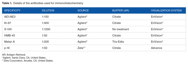
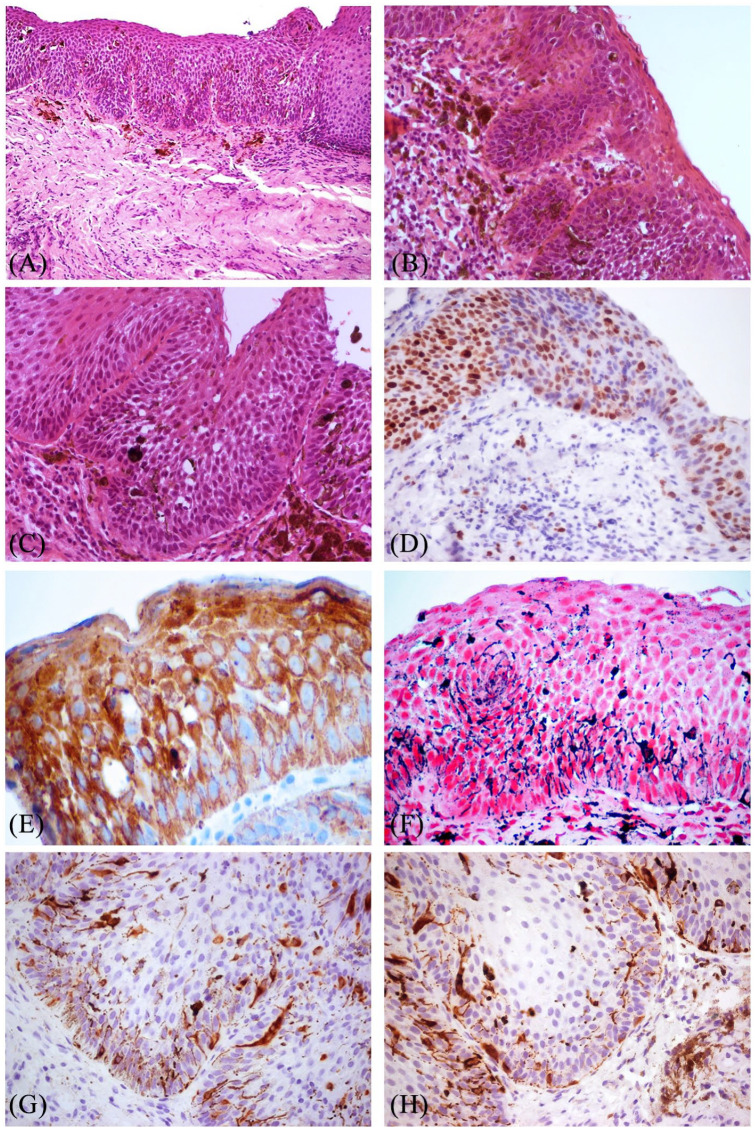
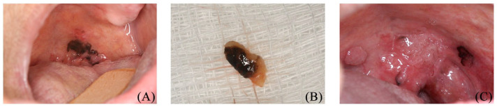
Pigmented lesions of the oral mucosa encompass several benign and malignant conditions that may be a matter of concern under both clinical and histopathological views. We reported a case of a 62-year-old woman, presenting with an asymptomatic, deeply pigmented lesion on the soft palate. On examination, it appeared asymmetrical, with irregular borders and an area of ulceration. A biopsy, taken to rule out melanoma, revealed a pigmented carcinoma in situ. Throughout the tumor thickness, numerous interspersed melanocytes were found that did not extend to neighboring epithelium. These were large, richly dendritic, and presented abundance of melanin granules and small nuclei. Mild melanin incontinence was found. Scanty transfer of pigment to dysplastic epithelial cells was found through Fontana Masson staining. On immunohistochemical analyses, there were pancytokeratin-stained tumor epithelial cells; increased cell proliferation throughout the entire thickness of the tumor was emphasized by Ki-67 immunomarking. P16 was negative. The dendritic cells were selectively stained for S-100, HMB45 and Melan A. Wide spectrum in situ hybridization for human papillomavirus (HPV) was negative. Unfortunately, following diagnosis, the patient refused any treatment option. Pigmented squamous cell carcinoma with melanocyte colonization must be taken into account in the differential diagnosis of pigmented lesions of the oral cavity.

 求助内容:
求助内容: 应助结果提醒方式:


