{"title":"肝肺泡包虫病的多模态影像学表现和弥散加权成像在其特征中的潜在作用。","authors":"Arshed H Parry, Abdul H Wani, Imza Feroz","doi":"10.5114/pjr.2020.101015","DOIUrl":null,"url":null,"abstract":"<p><strong>Purpose: </strong>To study the spectrum of imaging findings in hepatic alveolar echinococcosis (HAE) and to evaluate the potential role of diffusion-weighted imaging (DWI) in its characterisation.</p><p><strong>Material and methods: </strong>Two radiologists with more than seven years of experience retrospectively studied ultrasonography, computed tomography (CT), and magnetic resonance imaging (MRI) findings in 23 histopathologically proven cases of HAE with emphasis on the appearance and extent of disease. DWI characteristics of lesions were noted, and their apparent diffusion values (ADC) were calculated.</p><p><strong>Results: </strong>Ultrasonography features of HAE included heterogeneous, hyperechoic hepatic mass with or without calcification (<i>n</i> = 20), or heterogeneous mass with solid-cystic appearance (<i>n</i> = 2). CT revealed heterogeneous density infiltrative hepatic mass with no contrast enhancement in 19 patients or thick-walled cystic mass (<i>n</i> = 4). Following Kodama classification one type 1, six type 2, two type 3, eight type 4, and two type 5 lesions were identified on T2-weighted MRI. No enhancement was seen on post-contrast T1-weighted images. Mean ADC values were 1.74 ± 0.48 × 10<sup>-3</sup> mm<sup>2</sup>/s (range: 1.39 × 10<sup>-3</sup> mm<sup>2</sup>/s to 2.3 × 10<sup>-3</sup> mm<sup>2</sup>/s).</p><p><strong>Conclusions: </strong>HAE by virtue of its infiltrative growth pattern with a tendency to involve biliary, vascular, and extra hepatic structures can be easily misdiagnosed as malignant hepatic neoplasm. Knowledge of varied imaging appearances of HAE is essential to suspect the condition and to make an appropriate diagnosis. Diffusion-weighted imaging is a useful adjunct with relatively high diffusivity (high ADC values) suggesting diagnosis of alveolar hydatid.</p>","PeriodicalId":47128,"journal":{"name":"Polish Journal of Radiology","volume":"85 ","pages":"e613-e623"},"PeriodicalIF":0.9000,"publicationDate":"2020-11-10","publicationTypes":"Journal Article","fieldsOfStudy":null,"isOpenAccess":false,"openAccessPdf":"https://ftp.ncbi.nlm.nih.gov/pub/pmc/oa_pdf/bd/85/PJR-85-42479.PMC7757515.pdf","citationCount":"4","resultStr":"{\"title\":\"The spectrum of multimodality imaging findings in hepatic alveolar echinococcosis and the potential role of diffusion-weighted imaging in its characterisation.\",\"authors\":\"Arshed H Parry, Abdul H Wani, Imza Feroz\",\"doi\":\"10.5114/pjr.2020.101015\",\"DOIUrl\":null,\"url\":null,\"abstract\":\"<p><strong>Purpose: </strong>To study the spectrum of imaging findings in hepatic alveolar echinococcosis (HAE) and to evaluate the potential role of diffusion-weighted imaging (DWI) in its characterisation.</p><p><strong>Material and methods: </strong>Two radiologists with more than seven years of experience retrospectively studied ultrasonography, computed tomography (CT), and magnetic resonance imaging (MRI) findings in 23 histopathologically proven cases of HAE with emphasis on the appearance and extent of disease. DWI characteristics of lesions were noted, and their apparent diffusion values (ADC) were calculated.</p><p><strong>Results: </strong>Ultrasonography features of HAE included heterogeneous, hyperechoic hepatic mass with or without calcification (<i>n</i> = 20), or heterogeneous mass with solid-cystic appearance (<i>n</i> = 2). CT revealed heterogeneous density infiltrative hepatic mass with no contrast enhancement in 19 patients or thick-walled cystic mass (<i>n</i> = 4). Following Kodama classification one type 1, six type 2, two type 3, eight type 4, and two type 5 lesions were identified on T2-weighted MRI. No enhancement was seen on post-contrast T1-weighted images. Mean ADC values were 1.74 ± 0.48 × 10<sup>-3</sup> mm<sup>2</sup>/s (range: 1.39 × 10<sup>-3</sup> mm<sup>2</sup>/s to 2.3 × 10<sup>-3</sup> mm<sup>2</sup>/s).</p><p><strong>Conclusions: </strong>HAE by virtue of its infiltrative growth pattern with a tendency to involve biliary, vascular, and extra hepatic structures can be easily misdiagnosed as malignant hepatic neoplasm. Knowledge of varied imaging appearances of HAE is essential to suspect the condition and to make an appropriate diagnosis. Diffusion-weighted imaging is a useful adjunct with relatively high diffusivity (high ADC values) suggesting diagnosis of alveolar hydatid.</p>\",\"PeriodicalId\":47128,\"journal\":{\"name\":\"Polish Journal of Radiology\",\"volume\":\"85 \",\"pages\":\"e613-e623\"},\"PeriodicalIF\":0.9000,\"publicationDate\":\"2020-11-10\",\"publicationTypes\":\"Journal Article\",\"fieldsOfStudy\":null,\"isOpenAccess\":false,\"openAccessPdf\":\"https://ftp.ncbi.nlm.nih.gov/pub/pmc/oa_pdf/bd/85/PJR-85-42479.PMC7757515.pdf\",\"citationCount\":\"4\",\"resultStr\":null,\"platform\":\"Semanticscholar\",\"paperid\":null,\"PeriodicalName\":\"Polish Journal of Radiology\",\"FirstCategoryId\":\"1085\",\"ListUrlMain\":\"https://doi.org/10.5114/pjr.2020.101015\",\"RegionNum\":0,\"RegionCategory\":null,\"ArticlePicture\":[],\"TitleCN\":null,\"AbstractTextCN\":null,\"PMCID\":null,\"EPubDate\":\"2020/1/1 0:00:00\",\"PubModel\":\"eCollection\",\"JCR\":\"Q4\",\"JCRName\":\"RADIOLOGY, NUCLEAR MEDICINE & MEDICAL IMAGING\",\"Score\":null,\"Total\":0}","platform":"Semanticscholar","paperid":null,"PeriodicalName":"Polish Journal of Radiology","FirstCategoryId":"1085","ListUrlMain":"https://doi.org/10.5114/pjr.2020.101015","RegionNum":0,"RegionCategory":null,"ArticlePicture":[],"TitleCN":null,"AbstractTextCN":null,"PMCID":null,"EPubDate":"2020/1/1 0:00:00","PubModel":"eCollection","JCR":"Q4","JCRName":"RADIOLOGY, NUCLEAR MEDICINE & MEDICAL IMAGING","Score":null,"Total":0}
The spectrum of multimodality imaging findings in hepatic alveolar echinococcosis and the potential role of diffusion-weighted imaging in its characterisation.
Purpose: To study the spectrum of imaging findings in hepatic alveolar echinococcosis (HAE) and to evaluate the potential role of diffusion-weighted imaging (DWI) in its characterisation.
Material and methods: Two radiologists with more than seven years of experience retrospectively studied ultrasonography, computed tomography (CT), and magnetic resonance imaging (MRI) findings in 23 histopathologically proven cases of HAE with emphasis on the appearance and extent of disease. DWI characteristics of lesions were noted, and their apparent diffusion values (ADC) were calculated.
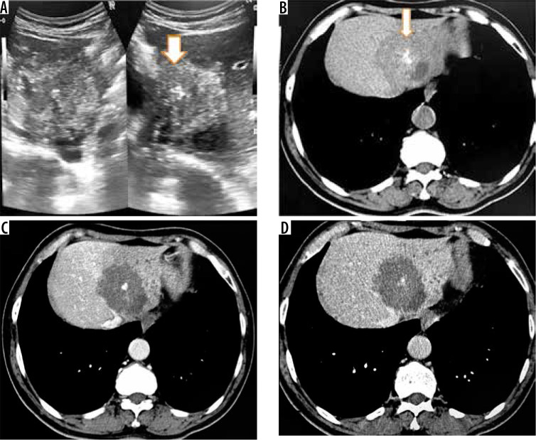
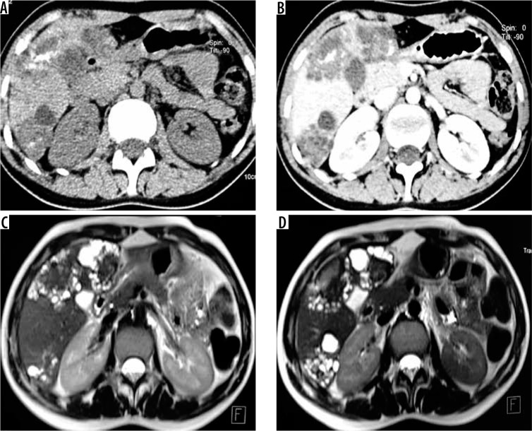
Results: Ultrasonography features of HAE included heterogeneous, hyperechoic hepatic mass with or without calcification (n = 20), or heterogeneous mass with solid-cystic appearance (n = 2). CT revealed heterogeneous density infiltrative hepatic mass with no contrast enhancement in 19 patients or thick-walled cystic mass (n = 4). Following Kodama classification one type 1, six type 2, two type 3, eight type 4, and two type 5 lesions were identified on T2-weighted MRI. No enhancement was seen on post-contrast T1-weighted images. Mean ADC values were 1.74 ± 0.48 × 10⁻³ mm2/s (range: 1.39 × 10⁻³ mm2/s to 2.3 × 10⁻³ mm2/s).
Conclusions: HAE by virtue of its infiltrative growth pattern with a tendency to involve biliary, vascular, and extra hepatic structures can be easily misdiagnosed as malignant hepatic neoplasm. Knowledge of varied imaging appearances of HAE is essential to suspect the condition and to make an appropriate diagnosis. Diffusion-weighted imaging is a useful adjunct with relatively high diffusivity (high ADC values) suggesting diagnosis of alveolar hydatid.


 求助内容:
求助内容: 应助结果提醒方式:


