Megan D. Lenardon , Prashant Sood , Helge C. Dorfmueller , Alistair J.P. Brown , Neil A.R. Gow
{"title":"白色念珠菌细胞壁的标量纳米结构分子,细胞和超微结构的分析和解释","authors":"Megan D. Lenardon , Prashant Sood , Helge C. Dorfmueller , Alistair J.P. Brown , Neil A.R. Gow","doi":"10.1016/j.tcsw.2020.100047","DOIUrl":null,"url":null,"abstract":"<div><p>Despite the importance of fungal cell walls as the principle determinant of fungal morphology and the defining element determining fungal interactions with other cells, few scalar models have been developed that reconcile chemical and microscopic attributes of its structure. The cell wall of the fungal pathogen <em>Candida albicans</em> is comprised of an amorphous inner skeletal layer of β(1,3)- and β(1,6)-glucan and chitin and an outer fibrillar layer thought to be dominated by highly mannosylated cell wall proteins. The architecture of these two layers can be resolved at the electron microscopy level, but the visualised structure of the wall has not yet been defined precisely in chemical terms. We have therefore examined the precise structure, location and molecular sizes of the cell wall components using transmission electron microscopy and tomography and tested predictions of the cell wall models using mutants and agents that perturb the normal cell wall structure. We demonstrate that the fibrils are comprised of a frond of <em>N</em>-linked outer chain mannans linked to a basal layer of GPI-proteins concentrated in the mid-wall region and that the non-elastic chitin microfibrils are cantilevered with sufficient lengths of non-fibrillar chitin and/or β-glucan to enable the chitin-glucan cage to flex, e.g. during morphogenesis and osmotic swelling. We present the first three-dimensional nano-scalar model of the <em>C. albicans</em> cell wall which can be used to test hypotheses relating to the structure–function relationships that underpin the pathobiology of this fungal pathogen.</p></div>","PeriodicalId":36539,"journal":{"name":"Cell Surface","volume":"6 ","pages":"Article 100047"},"PeriodicalIF":0.0000,"publicationDate":"2020-12-01","publicationTypes":"Journal Article","fieldsOfStudy":null,"isOpenAccess":false,"openAccessPdf":"https://sci-hub-pdf.com/10.1016/j.tcsw.2020.100047","citationCount":"31","resultStr":"{\"title\":\"Scalar nanostructure of the Candida albicans cell wall; a molecular, cellular and ultrastructural analysis and interpretation\",\"authors\":\"Megan D. Lenardon , Prashant Sood , Helge C. Dorfmueller , Alistair J.P. Brown , Neil A.R. Gow\",\"doi\":\"10.1016/j.tcsw.2020.100047\",\"DOIUrl\":null,\"url\":null,\"abstract\":\"<div><p>Despite the importance of fungal cell walls as the principle determinant of fungal morphology and the defining element determining fungal interactions with other cells, few scalar models have been developed that reconcile chemical and microscopic attributes of its structure. The cell wall of the fungal pathogen <em>Candida albicans</em> is comprised of an amorphous inner skeletal layer of β(1,3)- and β(1,6)-glucan and chitin and an outer fibrillar layer thought to be dominated by highly mannosylated cell wall proteins. The architecture of these two layers can be resolved at the electron microscopy level, but the visualised structure of the wall has not yet been defined precisely in chemical terms. We have therefore examined the precise structure, location and molecular sizes of the cell wall components using transmission electron microscopy and tomography and tested predictions of the cell wall models using mutants and agents that perturb the normal cell wall structure. We demonstrate that the fibrils are comprised of a frond of <em>N</em>-linked outer chain mannans linked to a basal layer of GPI-proteins concentrated in the mid-wall region and that the non-elastic chitin microfibrils are cantilevered with sufficient lengths of non-fibrillar chitin and/or β-glucan to enable the chitin-glucan cage to flex, e.g. during morphogenesis and osmotic swelling. We present the first three-dimensional nano-scalar model of the <em>C. albicans</em> cell wall which can be used to test hypotheses relating to the structure–function relationships that underpin the pathobiology of this fungal pathogen.</p></div>\",\"PeriodicalId\":36539,\"journal\":{\"name\":\"Cell Surface\",\"volume\":\"6 \",\"pages\":\"Article 100047\"},\"PeriodicalIF\":0.0000,\"publicationDate\":\"2020-12-01\",\"publicationTypes\":\"Journal Article\",\"fieldsOfStudy\":null,\"isOpenAccess\":false,\"openAccessPdf\":\"https://sci-hub-pdf.com/10.1016/j.tcsw.2020.100047\",\"citationCount\":\"31\",\"resultStr\":null,\"platform\":\"Semanticscholar\",\"paperid\":null,\"PeriodicalName\":\"Cell Surface\",\"FirstCategoryId\":\"1085\",\"ListUrlMain\":\"https://www.sciencedirect.com/science/article/pii/S2468233020300141\",\"RegionNum\":0,\"RegionCategory\":null,\"ArticlePicture\":[],\"TitleCN\":null,\"AbstractTextCN\":null,\"PMCID\":null,\"EPubDate\":\"\",\"PubModel\":\"\",\"JCR\":\"Q1\",\"JCRName\":\"Immunology and Microbiology\",\"Score\":null,\"Total\":0}","platform":"Semanticscholar","paperid":null,"PeriodicalName":"Cell Surface","FirstCategoryId":"1085","ListUrlMain":"https://www.sciencedirect.com/science/article/pii/S2468233020300141","RegionNum":0,"RegionCategory":null,"ArticlePicture":[],"TitleCN":null,"AbstractTextCN":null,"PMCID":null,"EPubDate":"","PubModel":"","JCR":"Q1","JCRName":"Immunology and Microbiology","Score":null,"Total":0}
Scalar nanostructure of the Candida albicans cell wall; a molecular, cellular and ultrastructural analysis and interpretation
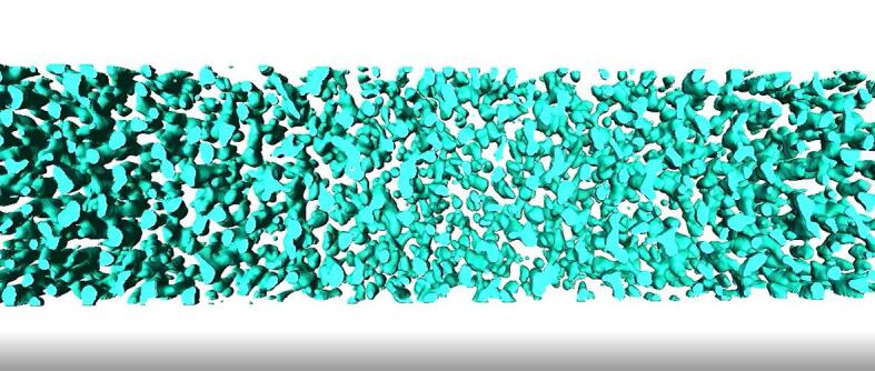
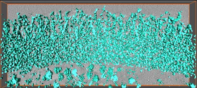
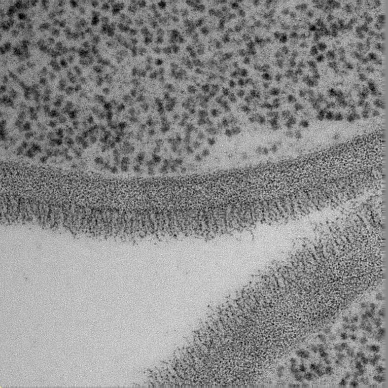
Despite the importance of fungal cell walls as the principle determinant of fungal morphology and the defining element determining fungal interactions with other cells, few scalar models have been developed that reconcile chemical and microscopic attributes of its structure. The cell wall of the fungal pathogen Candida albicans is comprised of an amorphous inner skeletal layer of β(1,3)- and β(1,6)-glucan and chitin and an outer fibrillar layer thought to be dominated by highly mannosylated cell wall proteins. The architecture of these two layers can be resolved at the electron microscopy level, but the visualised structure of the wall has not yet been defined precisely in chemical terms. We have therefore examined the precise structure, location and molecular sizes of the cell wall components using transmission electron microscopy and tomography and tested predictions of the cell wall models using mutants and agents that perturb the normal cell wall structure. We demonstrate that the fibrils are comprised of a frond of N-linked outer chain mannans linked to a basal layer of GPI-proteins concentrated in the mid-wall region and that the non-elastic chitin microfibrils are cantilevered with sufficient lengths of non-fibrillar chitin and/or β-glucan to enable the chitin-glucan cage to flex, e.g. during morphogenesis and osmotic swelling. We present the first three-dimensional nano-scalar model of the C. albicans cell wall which can be used to test hypotheses relating to the structure–function relationships that underpin the pathobiology of this fungal pathogen.

 求助内容:
求助内容: 应助结果提醒方式:


