Machiel P Ysebaert, Jessica P Johnson, Ghazanfar Abbas, Paulo Henrique Cavalcante, Rodney King, Masa Oikawa, Sarah Puchalski, Florent David
{"title":"关节镜下切除干扰马驹足滑车的孤立性骨软骨瘤。","authors":"Machiel P Ysebaert, Jessica P Johnson, Ghazanfar Abbas, Paulo Henrique Cavalcante, Rodney King, Masa Oikawa, Sarah Puchalski, Florent David","doi":"10.1111/vsu.13522","DOIUrl":null,"url":null,"abstract":"<p><strong>Objective: </strong>To report the diagnostics, surgical treatment, and outcome of a juvenile foal with solitary osteochondroma (SO) interfering with the podotrochlear apparatus.</p><p><strong>Study design: </strong>Case report ANIMAL: One 36-day-old Arabian colt.</p><p><strong>Methods: </strong>Clinical, radiographic, ultrasonographic, computed tomographic, and histopathologic examinations were required to characterize and treat an SO located at the palmar aspect of the diaphysis of the second phalanx of the left forelimb. This SO caused severe distal interphalangeal joint (DIPJ) inflammation, marked interference with the podotrochlear apparatus, and associated lameness.</p><p><strong>Results: </strong>Despite the small size of the foal's foot, complete resection of the SO was possible via palmar DIPJ arthroscopy by using motorized equipment. Full resolution of the lameness was achieved within 3 months of surgery.</p><p><strong>Conclusion: </strong>Atypical SO located on the palmar aspect of the second phalanx can cause marked nonseptic inflammation of the DIPJ and interference with the podotrochlear apparatus and should be considered among the differential diagnoses for severe lameness in juvenile foals. Arthroscopic resection of the SO led to an excellent outcome.</p>","PeriodicalId":123280,"journal":{"name":"Veterinary surgery : VS","volume":" ","pages":"O128-O137"},"PeriodicalIF":0.0000,"publicationDate":"2021-07-01","publicationTypes":"Journal Article","fieldsOfStudy":null,"isOpenAccess":false,"openAccessPdf":"https://sci-hub-pdf.com/10.1111/vsu.13522","citationCount":"0","resultStr":"{\"title\":\"Arthroscopic removal of a solitary osteochondroma interfering with the podotrochlear apparatus in a foal.\",\"authors\":\"Machiel P Ysebaert, Jessica P Johnson, Ghazanfar Abbas, Paulo Henrique Cavalcante, Rodney King, Masa Oikawa, Sarah Puchalski, Florent David\",\"doi\":\"10.1111/vsu.13522\",\"DOIUrl\":null,\"url\":null,\"abstract\":\"<p><strong>Objective: </strong>To report the diagnostics, surgical treatment, and outcome of a juvenile foal with solitary osteochondroma (SO) interfering with the podotrochlear apparatus.</p><p><strong>Study design: </strong>Case report ANIMAL: One 36-day-old Arabian colt.</p><p><strong>Methods: </strong>Clinical, radiographic, ultrasonographic, computed tomographic, and histopathologic examinations were required to characterize and treat an SO located at the palmar aspect of the diaphysis of the second phalanx of the left forelimb. This SO caused severe distal interphalangeal joint (DIPJ) inflammation, marked interference with the podotrochlear apparatus, and associated lameness.</p><p><strong>Results: </strong>Despite the small size of the foal's foot, complete resection of the SO was possible via palmar DIPJ arthroscopy by using motorized equipment. Full resolution of the lameness was achieved within 3 months of surgery.</p><p><strong>Conclusion: </strong>Atypical SO located on the palmar aspect of the second phalanx can cause marked nonseptic inflammation of the DIPJ and interference with the podotrochlear apparatus and should be considered among the differential diagnoses for severe lameness in juvenile foals. Arthroscopic resection of the SO led to an excellent outcome.</p>\",\"PeriodicalId\":123280,\"journal\":{\"name\":\"Veterinary surgery : VS\",\"volume\":\" \",\"pages\":\"O128-O137\"},\"PeriodicalIF\":0.0000,\"publicationDate\":\"2021-07-01\",\"publicationTypes\":\"Journal Article\",\"fieldsOfStudy\":null,\"isOpenAccess\":false,\"openAccessPdf\":\"https://sci-hub-pdf.com/10.1111/vsu.13522\",\"citationCount\":\"0\",\"resultStr\":null,\"platform\":\"Semanticscholar\",\"paperid\":null,\"PeriodicalName\":\"Veterinary surgery : VS\",\"FirstCategoryId\":\"97\",\"ListUrlMain\":\"https://doi.org/10.1111/vsu.13522\",\"RegionNum\":0,\"RegionCategory\":null,\"ArticlePicture\":[],\"TitleCN\":null,\"AbstractTextCN\":null,\"PMCID\":null,\"EPubDate\":\"2020/10/14 0:00:00\",\"PubModel\":\"Epub\",\"JCR\":\"\",\"JCRName\":\"\",\"Score\":null,\"Total\":0}","platform":"Semanticscholar","paperid":null,"PeriodicalName":"Veterinary surgery : VS","FirstCategoryId":"97","ListUrlMain":"https://doi.org/10.1111/vsu.13522","RegionNum":0,"RegionCategory":null,"ArticlePicture":[],"TitleCN":null,"AbstractTextCN":null,"PMCID":null,"EPubDate":"2020/10/14 0:00:00","PubModel":"Epub","JCR":"","JCRName":"","Score":null,"Total":0}
Arthroscopic removal of a solitary osteochondroma interfering with the podotrochlear apparatus in a foal.
Objective: To report the diagnostics, surgical treatment, and outcome of a juvenile foal with solitary osteochondroma (SO) interfering with the podotrochlear apparatus.
Study design: Case report ANIMAL: One 36-day-old Arabian colt.
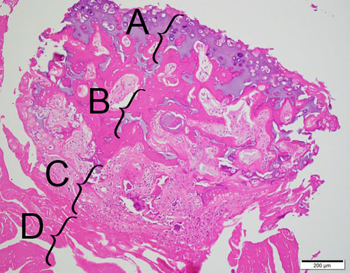
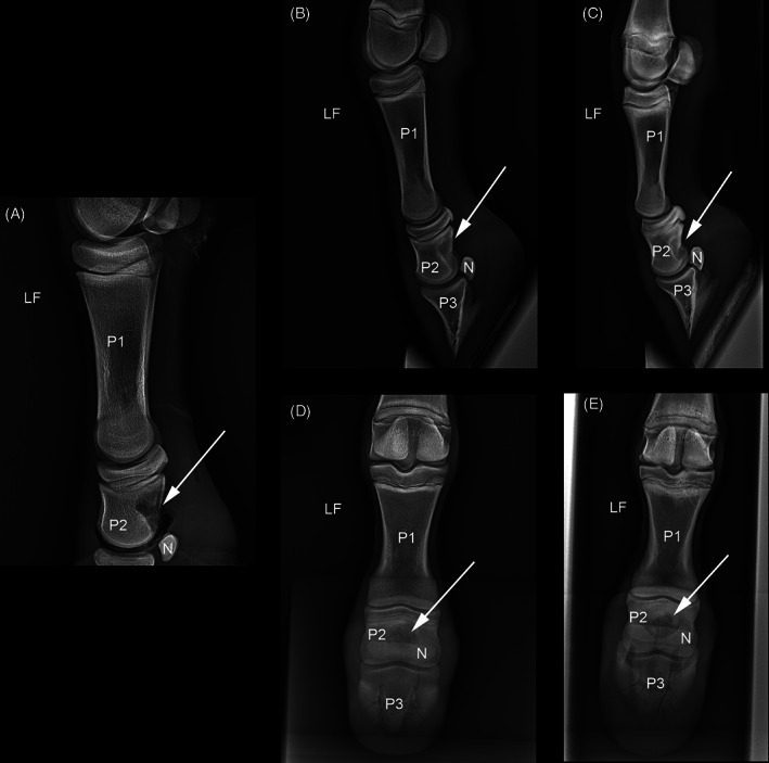
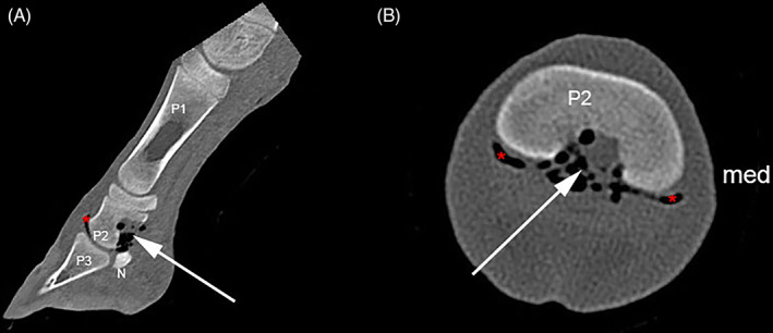
Methods: Clinical, radiographic, ultrasonographic, computed tomographic, and histopathologic examinations were required to characterize and treat an SO located at the palmar aspect of the diaphysis of the second phalanx of the left forelimb. This SO caused severe distal interphalangeal joint (DIPJ) inflammation, marked interference with the podotrochlear apparatus, and associated lameness.
Results: Despite the small size of the foal's foot, complete resection of the SO was possible via palmar DIPJ arthroscopy by using motorized equipment. Full resolution of the lameness was achieved within 3 months of surgery.
Conclusion: Atypical SO located on the palmar aspect of the second phalanx can cause marked nonseptic inflammation of the DIPJ and interference with the podotrochlear apparatus and should be considered among the differential diagnoses for severe lameness in juvenile foals. Arthroscopic resection of the SO led to an excellent outcome.

 求助内容:
求助内容: 应助结果提醒方式:


