Keno K Bressem, Lisa C Adams, Jakob Albrecht, Antonie Petersen, Hans-Martin Thieß, Alexandra Niehues, Stefan M Niehues, Janis L Vahldiek
{"title":"肺密度与COVID-19的严重程度有关吗?","authors":"Keno K Bressem, Lisa C Adams, Jakob Albrecht, Antonie Petersen, Hans-Martin Thieß, Alexandra Niehues, Stefan M Niehues, Janis L Vahldiek","doi":"10.5114/pjr.2020.100788","DOIUrl":null,"url":null,"abstract":"<p><strong>Purpose: </strong>Emphysema and chronic obstructive lung disease were previously identified as major risk factors for severe disease progression in COVID-19. Computed tomography (CT)-based lung-density analysis offers a fast, reliable, and quantitative assessment of lung density. Therefore, we aimed to assess the benefit of CT-based lung density measurements to predict possible severe disease progression in COVID-19.</p><p><strong>Material and methods: </strong>Thirty COVID-19-positive patients were included in this retrospective study. Lung density was quantified based on routinely acquired chest CTs. Presence of COVID-19 was confirmed by reverse transcription polymerase chain reaction (RT-PCR). Wilcoxon test was used to compare two groups of patients. A multivariate regression analysis, adjusted for age and sex, was employed to model the relative increase of risk for severe disease, depending on the measured densities.</p><p><strong>Results: </strong>Intensive care unit (ICU) patients or patients requiring mechanical ventilation showed a lower proportion of medium- and low-density lung volume compared to patients on the normal ward, but a significantly larger volume of high-density lung volume (12.26 dl IQR 4.65 dl vs. 7.51 dl vs. IQR 5.39 dl, <i>p</i> = 0.039). In multivariate regression analysis, high-density lung volume was identified as a significant predictor of severe disease.</p><p><strong>Conclusions: </strong>The amount of high-density lung tissue showed a significant association with severe COVID-19, with odds ratios of 1.42 (95% CI: 1.09-2.00) and 1.37 (95% CI: 1.03-2.11) for requiring intensive care and mechanical ventilation, respectively. Acknowledging our small sample size as an important limitation; our study might thus suggest that high-density lung tissue could serve as a possible predictor of severe COVID-19.</p>","PeriodicalId":47128,"journal":{"name":"Polish Journal of Radiology","volume":"85 ","pages":"e600-e606"},"PeriodicalIF":0.9000,"publicationDate":"2020-10-30","publicationTypes":"Journal Article","fieldsOfStudy":null,"isOpenAccess":false,"openAccessPdf":"https://ftp.ncbi.nlm.nih.gov/pub/pmc/oa_pdf/61/f0/PJR-85-42392.PMC7654311.pdf","citationCount":"6","resultStr":"{\"title\":\"Is lung density associated with severity of COVID-19?\",\"authors\":\"Keno K Bressem, Lisa C Adams, Jakob Albrecht, Antonie Petersen, Hans-Martin Thieß, Alexandra Niehues, Stefan M Niehues, Janis L Vahldiek\",\"doi\":\"10.5114/pjr.2020.100788\",\"DOIUrl\":null,\"url\":null,\"abstract\":\"<p><strong>Purpose: </strong>Emphysema and chronic obstructive lung disease were previously identified as major risk factors for severe disease progression in COVID-19. Computed tomography (CT)-based lung-density analysis offers a fast, reliable, and quantitative assessment of lung density. Therefore, we aimed to assess the benefit of CT-based lung density measurements to predict possible severe disease progression in COVID-19.</p><p><strong>Material and methods: </strong>Thirty COVID-19-positive patients were included in this retrospective study. Lung density was quantified based on routinely acquired chest CTs. Presence of COVID-19 was confirmed by reverse transcription polymerase chain reaction (RT-PCR). Wilcoxon test was used to compare two groups of patients. A multivariate regression analysis, adjusted for age and sex, was employed to model the relative increase of risk for severe disease, depending on the measured densities.</p><p><strong>Results: </strong>Intensive care unit (ICU) patients or patients requiring mechanical ventilation showed a lower proportion of medium- and low-density lung volume compared to patients on the normal ward, but a significantly larger volume of high-density lung volume (12.26 dl IQR 4.65 dl vs. 7.51 dl vs. IQR 5.39 dl, <i>p</i> = 0.039). In multivariate regression analysis, high-density lung volume was identified as a significant predictor of severe disease.</p><p><strong>Conclusions: </strong>The amount of high-density lung tissue showed a significant association with severe COVID-19, with odds ratios of 1.42 (95% CI: 1.09-2.00) and 1.37 (95% CI: 1.03-2.11) for requiring intensive care and mechanical ventilation, respectively. Acknowledging our small sample size as an important limitation; our study might thus suggest that high-density lung tissue could serve as a possible predictor of severe COVID-19.</p>\",\"PeriodicalId\":47128,\"journal\":{\"name\":\"Polish Journal of Radiology\",\"volume\":\"85 \",\"pages\":\"e600-e606\"},\"PeriodicalIF\":0.9000,\"publicationDate\":\"2020-10-30\",\"publicationTypes\":\"Journal Article\",\"fieldsOfStudy\":null,\"isOpenAccess\":false,\"openAccessPdf\":\"https://ftp.ncbi.nlm.nih.gov/pub/pmc/oa_pdf/61/f0/PJR-85-42392.PMC7654311.pdf\",\"citationCount\":\"6\",\"resultStr\":null,\"platform\":\"Semanticscholar\",\"paperid\":null,\"PeriodicalName\":\"Polish Journal of Radiology\",\"FirstCategoryId\":\"1085\",\"ListUrlMain\":\"https://doi.org/10.5114/pjr.2020.100788\",\"RegionNum\":0,\"RegionCategory\":null,\"ArticlePicture\":[],\"TitleCN\":null,\"AbstractTextCN\":null,\"PMCID\":null,\"EPubDate\":\"2020/1/1 0:00:00\",\"PubModel\":\"eCollection\",\"JCR\":\"Q4\",\"JCRName\":\"RADIOLOGY, NUCLEAR MEDICINE & MEDICAL IMAGING\",\"Score\":null,\"Total\":0}","platform":"Semanticscholar","paperid":null,"PeriodicalName":"Polish Journal of Radiology","FirstCategoryId":"1085","ListUrlMain":"https://doi.org/10.5114/pjr.2020.100788","RegionNum":0,"RegionCategory":null,"ArticlePicture":[],"TitleCN":null,"AbstractTextCN":null,"PMCID":null,"EPubDate":"2020/1/1 0:00:00","PubModel":"eCollection","JCR":"Q4","JCRName":"RADIOLOGY, NUCLEAR MEDICINE & MEDICAL IMAGING","Score":null,"Total":0}
引用次数: 6
摘要
目的:肺气肿和慢性阻塞性肺疾病之前被确定为COVID-19严重疾病进展的主要危险因素。基于计算机断层扫描(CT)的肺密度分析提供了一种快速、可靠和定量的肺密度评估。因此,我们旨在评估基于ct的肺密度测量在预测COVID-19可能的严重疾病进展方面的益处。材料与方法:回顾性研究30例covid -19阳性患者。肺密度根据常规胸部ct进行量化。逆转录聚合酶链反应(RT-PCR)证实存在COVID-19。采用Wilcoxon检验对两组患者进行比较。根据测量的密度,采用多变量回归分析,调整年龄和性别,对严重疾病风险的相对增加进行建模。结果:重症监护病房(ICU)患者或需要机械通气的患者中、低密度肺容量比例低于普通病房患者,但高密度肺容量明显高于普通病房患者(12.26 dl IQR 4.65 dl vs. 7.51 dl vs. 5.39 dl, p = 0.039)。在多变量回归分析中,高密度肺容量被确定为严重疾病的重要预测因子。结论:高密度肺组织的数量与重症COVID-19相关,需要重症监护和机械通气的优势比分别为1.42 (95% CI: 1.09-2.00)和1.37 (95% CI: 1.03-2.11)。承认我们的小样本量是一个重要的限制;因此,我们的研究可能表明高密度肺组织可以作为严重COVID-19的可能预测指标。
Is lung density associated with severity of COVID-19?
Purpose: Emphysema and chronic obstructive lung disease were previously identified as major risk factors for severe disease progression in COVID-19. Computed tomography (CT)-based lung-density analysis offers a fast, reliable, and quantitative assessment of lung density. Therefore, we aimed to assess the benefit of CT-based lung density measurements to predict possible severe disease progression in COVID-19.
Material and methods: Thirty COVID-19-positive patients were included in this retrospective study. Lung density was quantified based on routinely acquired chest CTs. Presence of COVID-19 was confirmed by reverse transcription polymerase chain reaction (RT-PCR). Wilcoxon test was used to compare two groups of patients. A multivariate regression analysis, adjusted for age and sex, was employed to model the relative increase of risk for severe disease, depending on the measured densities.
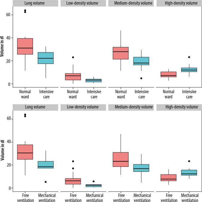
Results: Intensive care unit (ICU) patients or patients requiring mechanical ventilation showed a lower proportion of medium- and low-density lung volume compared to patients on the normal ward, but a significantly larger volume of high-density lung volume (12.26 dl IQR 4.65 dl vs. 7.51 dl vs. IQR 5.39 dl, p = 0.039). In multivariate regression analysis, high-density lung volume was identified as a significant predictor of severe disease.
Conclusions: The amount of high-density lung tissue showed a significant association with severe COVID-19, with odds ratios of 1.42 (95% CI: 1.09-2.00) and 1.37 (95% CI: 1.03-2.11) for requiring intensive care and mechanical ventilation, respectively. Acknowledging our small sample size as an important limitation; our study might thus suggest that high-density lung tissue could serve as a possible predictor of severe COVID-19.

 求助内容:
求助内容: 应助结果提醒方式:


