{"title":"用3特斯拉磁共振成像对健康和患病耳耳咽管软骨的比较评价。","authors":"Nevin Aydın, Suzan Saylısoy, Baki Adapınar, Didem Arslantas","doi":"10.5114/pjr.2020.99756","DOIUrl":null,"url":null,"abstract":"<p><strong>Purpose: </strong>The purpose of this study was to prospectively assess the Eustachian tube (ET) cartilage using 3 Tesla (3T) magnetic resonance imaging (MRI) and compare the results between healthy ears and those with a middle ear disease.</p><p><strong>Material and methods: </strong>The study included 56 ears with a middle ear disease as the patient group and 100 ears without a middle ear disease as the control group. The patients' age ranged from 18 to 65 years. The axial three-dimensional (3D) multiple echo recombined gradient echo (MERGE) sequence and oblique parasagittal planes were obtained. Visualisation of the ET cartilage was assessed on the MR images using a three-point numerical rating score. In the axial plane, the ET lumen's diameter was measured from the mid-portion of the cartilage.</p><p><strong>Results: </strong>There was no significant difference between the patient group and the control group according to patients' age and gender, and the medial laminal thickness of the ET cartilage. In the patient group, the diameter of the ET cartilage was significantly smaller than in the control group. The ET lumen diameter was significantly lower according to each of the three scoring systems.</p><p><strong>Conclusions: </strong>3T MRI provides an evaluation of the ET cartilage and isthmus level, which are small but important anatomical localisations and surgical landmarks. MR imaging has the potential to provide essential information on ET prior to new surgical treatments, such as balloon dilation for middle ear diseases.</p>","PeriodicalId":47128,"journal":{"name":"Polish Journal of Radiology","volume":"85 ","pages":"e581-e585"},"PeriodicalIF":0.9000,"publicationDate":"2020-10-15","publicationTypes":"Journal Article","fieldsOfStudy":null,"isOpenAccess":false,"openAccessPdf":"https://ftp.ncbi.nlm.nih.gov/pub/pmc/oa_pdf/b5/9f/PJR-85-42015.PMC7654315.pdf","citationCount":"1","resultStr":"{\"title\":\"A comparative evaluation of the Eustachian tube cartilage between healthy and diseased ears using a 3 Tesla MRI.\",\"authors\":\"Nevin Aydın, Suzan Saylısoy, Baki Adapınar, Didem Arslantas\",\"doi\":\"10.5114/pjr.2020.99756\",\"DOIUrl\":null,\"url\":null,\"abstract\":\"<p><strong>Purpose: </strong>The purpose of this study was to prospectively assess the Eustachian tube (ET) cartilage using 3 Tesla (3T) magnetic resonance imaging (MRI) and compare the results between healthy ears and those with a middle ear disease.</p><p><strong>Material and methods: </strong>The study included 56 ears with a middle ear disease as the patient group and 100 ears without a middle ear disease as the control group. The patients' age ranged from 18 to 65 years. The axial three-dimensional (3D) multiple echo recombined gradient echo (MERGE) sequence and oblique parasagittal planes were obtained. Visualisation of the ET cartilage was assessed on the MR images using a three-point numerical rating score. In the axial plane, the ET lumen's diameter was measured from the mid-portion of the cartilage.</p><p><strong>Results: </strong>There was no significant difference between the patient group and the control group according to patients' age and gender, and the medial laminal thickness of the ET cartilage. In the patient group, the diameter of the ET cartilage was significantly smaller than in the control group. The ET lumen diameter was significantly lower according to each of the three scoring systems.</p><p><strong>Conclusions: </strong>3T MRI provides an evaluation of the ET cartilage and isthmus level, which are small but important anatomical localisations and surgical landmarks. MR imaging has the potential to provide essential information on ET prior to new surgical treatments, such as balloon dilation for middle ear diseases.</p>\",\"PeriodicalId\":47128,\"journal\":{\"name\":\"Polish Journal of Radiology\",\"volume\":\"85 \",\"pages\":\"e581-e585\"},\"PeriodicalIF\":0.9000,\"publicationDate\":\"2020-10-15\",\"publicationTypes\":\"Journal Article\",\"fieldsOfStudy\":null,\"isOpenAccess\":false,\"openAccessPdf\":\"https://ftp.ncbi.nlm.nih.gov/pub/pmc/oa_pdf/b5/9f/PJR-85-42015.PMC7654315.pdf\",\"citationCount\":\"1\",\"resultStr\":null,\"platform\":\"Semanticscholar\",\"paperid\":null,\"PeriodicalName\":\"Polish Journal of Radiology\",\"FirstCategoryId\":\"1085\",\"ListUrlMain\":\"https://doi.org/10.5114/pjr.2020.99756\",\"RegionNum\":0,\"RegionCategory\":null,\"ArticlePicture\":[],\"TitleCN\":null,\"AbstractTextCN\":null,\"PMCID\":null,\"EPubDate\":\"2020/1/1 0:00:00\",\"PubModel\":\"eCollection\",\"JCR\":\"Q4\",\"JCRName\":\"RADIOLOGY, NUCLEAR MEDICINE & MEDICAL IMAGING\",\"Score\":null,\"Total\":0}","platform":"Semanticscholar","paperid":null,"PeriodicalName":"Polish Journal of Radiology","FirstCategoryId":"1085","ListUrlMain":"https://doi.org/10.5114/pjr.2020.99756","RegionNum":0,"RegionCategory":null,"ArticlePicture":[],"TitleCN":null,"AbstractTextCN":null,"PMCID":null,"EPubDate":"2020/1/1 0:00:00","PubModel":"eCollection","JCR":"Q4","JCRName":"RADIOLOGY, NUCLEAR MEDICINE & MEDICAL IMAGING","Score":null,"Total":0}
A comparative evaluation of the Eustachian tube cartilage between healthy and diseased ears using a 3 Tesla MRI.
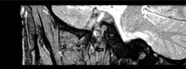
Purpose: The purpose of this study was to prospectively assess the Eustachian tube (ET) cartilage using 3 Tesla (3T) magnetic resonance imaging (MRI) and compare the results between healthy ears and those with a middle ear disease.
Material and methods: The study included 56 ears with a middle ear disease as the patient group and 100 ears without a middle ear disease as the control group. The patients' age ranged from 18 to 65 years. The axial three-dimensional (3D) multiple echo recombined gradient echo (MERGE) sequence and oblique parasagittal planes were obtained. Visualisation of the ET cartilage was assessed on the MR images using a three-point numerical rating score. In the axial plane, the ET lumen's diameter was measured from the mid-portion of the cartilage.
Results: There was no significant difference between the patient group and the control group according to patients' age and gender, and the medial laminal thickness of the ET cartilage. In the patient group, the diameter of the ET cartilage was significantly smaller than in the control group. The ET lumen diameter was significantly lower according to each of the three scoring systems.
Conclusions: 3T MRI provides an evaluation of the ET cartilage and isthmus level, which are small but important anatomical localisations and surgical landmarks. MR imaging has the potential to provide essential information on ET prior to new surgical treatments, such as balloon dilation for middle ear diseases.


 求助内容:
求助内容: 应助结果提醒方式:


