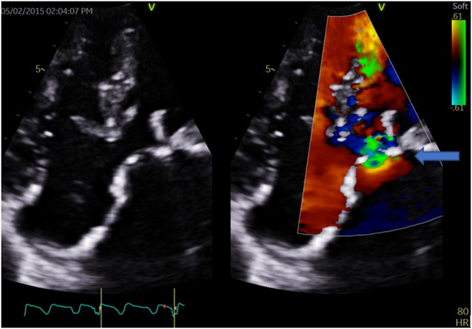
成人三房心或左心房分裂表现为二尖瓣狭窄。

IF 3.2 Q2 CARDIAC & CARDIOVASCULAR SYSTEMS
Alfonso Pecoraro, Jacques Janson, Jacob Daniel Cilliers
{"title":"成人三房心或左心房分裂表现为二尖瓣狭窄。","authors":"Alfonso Pecoraro, Jacques Janson, Jacob Daniel Cilliers","doi":"10.1530/ERP-20-0016","DOIUrl":null,"url":null,"abstract":"A 26-year-old male patient presented to the hospital with a 2-month history of progressive dyspnoea. He denied chest pain, coughing, orthopnoea, paroxysmal nocturnal dyspnoea, syncope or pre-syncope. He had no other significant comorbidities and he was not on any chronic medication. His cardiovascular examination revealed an undisplaced apex beat with a parasternal heave and a loud second heart sound (P2) suggestive of pulmonary hypertension. On auscultation, the first heart sound was normal with a loud second heart sound and a diastolic rumble. Given these findings, a clinical diagnosis of severe mitral stenosis with pulmonary hypertension was made. ECG was atypical for mitral stenosis, it revealed a dilated LA with left axis deviation due to a left anterior hemiblock. No features of right ventricular hypertrophy was noted. To our surprise, echocardiographic evaluation (Figs 1 and 2) revealed a primum ASD with the normal function of the left and right atrioventricular (AV) valve. A left-sided supra-valvular ridge or divided left atrium was identified with peak and mean gradients of 43/21 mmHg, respectively (Fig. 3). Video 1 is an apical four-chamber view of the defect, pre-operatively. -20-0016 ID: 20-0016","PeriodicalId":45749,"journal":{"name":"Echo Research and Practice","volume":null,"pages":null},"PeriodicalIF":3.2000,"publicationDate":"2020-12-01","publicationTypes":"Journal Article","fieldsOfStudy":null,"isOpenAccess":false,"openAccessPdf":"https://ftp.ncbi.nlm.nih.gov/pub/pmc/oa_pdf/da/80/ERP-20-0016.PMC7576639.pdf","citationCount":"0","resultStr":"{\"title\":\"Cor triatriatrum or divided left atrium presenting as mitral stenosis in an adult patient.\",\"authors\":\"Alfonso Pecoraro, Jacques Janson, Jacob Daniel Cilliers\",\"doi\":\"10.1530/ERP-20-0016\",\"DOIUrl\":null,\"url\":null,\"abstract\":\"A 26-year-old male patient presented to the hospital with a 2-month history of progressive dyspnoea. He denied chest pain, coughing, orthopnoea, paroxysmal nocturnal dyspnoea, syncope or pre-syncope. He had no other significant comorbidities and he was not on any chronic medication. His cardiovascular examination revealed an undisplaced apex beat with a parasternal heave and a loud second heart sound (P2) suggestive of pulmonary hypertension. On auscultation, the first heart sound was normal with a loud second heart sound and a diastolic rumble. Given these findings, a clinical diagnosis of severe mitral stenosis with pulmonary hypertension was made. ECG was atypical for mitral stenosis, it revealed a dilated LA with left axis deviation due to a left anterior hemiblock. No features of right ventricular hypertrophy was noted. To our surprise, echocardiographic evaluation (Figs 1 and 2) revealed a primum ASD with the normal function of the left and right atrioventricular (AV) valve. A left-sided supra-valvular ridge or divided left atrium was identified with peak and mean gradients of 43/21 mmHg, respectively (Fig. 3). Video 1 is an apical four-chamber view of the defect, pre-operatively. -20-0016 ID: 20-0016\",\"PeriodicalId\":45749,\"journal\":{\"name\":\"Echo Research and Practice\",\"volume\":null,\"pages\":null},\"PeriodicalIF\":3.2000,\"publicationDate\":\"2020-12-01\",\"publicationTypes\":\"Journal Article\",\"fieldsOfStudy\":null,\"isOpenAccess\":false,\"openAccessPdf\":\"https://ftp.ncbi.nlm.nih.gov/pub/pmc/oa_pdf/da/80/ERP-20-0016.PMC7576639.pdf\",\"citationCount\":\"0\",\"resultStr\":null,\"platform\":\"Semanticscholar\",\"paperid\":null,\"PeriodicalName\":\"Echo Research and Practice\",\"FirstCategoryId\":\"1085\",\"ListUrlMain\":\"https://doi.org/10.1530/ERP-20-0016\",\"RegionNum\":0,\"RegionCategory\":null,\"ArticlePicture\":[],\"TitleCN\":null,\"AbstractTextCN\":null,\"PMCID\":null,\"EPubDate\":\"\",\"PubModel\":\"\",\"JCR\":\"Q2\",\"JCRName\":\"CARDIAC & CARDIOVASCULAR SYSTEMS\",\"Score\":null,\"Total\":0}","platform":"Semanticscholar","paperid":null,"PeriodicalName":"Echo Research and Practice","FirstCategoryId":"1085","ListUrlMain":"https://doi.org/10.1530/ERP-20-0016","RegionNum":0,"RegionCategory":null,"ArticlePicture":[],"TitleCN":null,"AbstractTextCN":null,"PMCID":null,"EPubDate":"","PubModel":"","JCR":"Q2","JCRName":"CARDIAC & CARDIOVASCULAR SYSTEMS","Score":null,"Total":0}
引用次数: 0

摘要

本文章由计算机程序翻译,如有差异,请以英文原文为准。
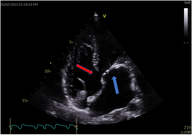
Cor triatriatrum or divided left atrium presenting as mitral stenosis in an adult patient.

Cor triatriatrum or divided left atrium presenting as mitral stenosis in an adult patient.

Cor triatriatrum or divided left atrium presenting as mitral stenosis in an adult patient.

Cor triatriatrum or divided left atrium presenting as mitral stenosis in an adult patient.
A 26-year-old male patient presented to the hospital with a 2-month history of progressive dyspnoea. He denied chest pain, coughing, orthopnoea, paroxysmal nocturnal dyspnoea, syncope or pre-syncope. He had no other significant comorbidities and he was not on any chronic medication. His cardiovascular examination revealed an undisplaced apex beat with a parasternal heave and a loud second heart sound (P2) suggestive of pulmonary hypertension. On auscultation, the first heart sound was normal with a loud second heart sound and a diastolic rumble. Given these findings, a clinical diagnosis of severe mitral stenosis with pulmonary hypertension was made. ECG was atypical for mitral stenosis, it revealed a dilated LA with left axis deviation due to a left anterior hemiblock. No features of right ventricular hypertrophy was noted. To our surprise, echocardiographic evaluation (Figs 1 and 2) revealed a primum ASD with the normal function of the left and right atrioventricular (AV) valve. A left-sided supra-valvular ridge or divided left atrium was identified with peak and mean gradients of 43/21 mmHg, respectively (Fig. 3). Video 1 is an apical four-chamber view of the defect, pre-operatively. -20-0016 ID: 20-0016
求助全文
通过发布文献求助,成功后即可免费获取论文全文。 去求助
来源期刊
Echo Research and Practice
Echo Research and Practice CARDIAC & CARDIOVASCULAR SYSTEMS-
CiteScore
6.70
自引率
12.70%
发文量
11
审稿时长
8 weeks
期刊介绍: Echo Research and Practice aims to be the premier international journal for physicians, sonographers, nurses and other allied health professionals practising echocardiography and other cardiac imaging modalities. This open-access journal publishes quality clinical and basic research, reviews, videos, education materials and selected high-interest case reports and videos across all echocardiography modalities and disciplines, including paediatrics, anaesthetics, general practice, acute medicine and intensive care. Multi-modality studies primarily featuring the use of cardiac ultrasound in clinical practice, in association with Cardiac Computed Tomography, Cardiovascular Magnetic Resonance or Nuclear Cardiology are of interest. Topics include, but are not limited to: 2D echocardiography 3D echocardiography Comparative imaging techniques – CCT, CMR and Nuclear Cardiology Congenital heart disease, including foetal echocardiography Contrast echocardiography Critical care echocardiography Deformation imaging Doppler echocardiography Interventional echocardiography Intracardiac echocardiography Intraoperative echocardiography Prosthetic valves Stress echocardiography Technical innovations Transoesophageal echocardiography Valve disease.
×
引用
GB/T 7714-2015
复制
MLA
复制
APA
复制
导出至
BibTeX EndNote RefMan NoteFirst NoteExpress
×
提示
您的信息不完整,为了账户安全,请先补充。
现在去补充
×
提示
您因"违规操作"
具体请查看互助需知
我知道了
×
提示
确定
请完成安全验证×
copy
已复制链接
快去分享给好友吧!
我知道了
右上角分享
点击右上角分享
0
联系我们:info@booksci.cn Book学术提供免费学术资源搜索服务,方便国内外学者检索中英文文献。致力于提供最便捷和优质的服务体验。 Copyright © 2023 布克学术 All rights reserved.
京ICP备2023020795号-1
ghs 京公网安备 11010802042870号
Book学术文献互助
Book学术文献互助群
群 号:481959085
Book学术官方微信