Dong Kyu Kim, Ji Min Lee, Seon Yeong Heo, Jong Pil Jung, Chang Ryul Park, Yong Jik Lee, Sang Cjeol Lee, Su Kyung Hwang, Gwan Sic Kim
{"title":"完全性倒位患者的急性A型主动脉夹层。","authors":"Dong Kyu Kim, Ji Min Lee, Seon Yeong Heo, Jong Pil Jung, Chang Ryul Park, Yong Jik Lee, Sang Cjeol Lee, Su Kyung Hwang, Gwan Sic Kim","doi":"10.5090/kjtcs.20.006","DOIUrl":null,"url":null,"abstract":"<p><p>We describe the occurrence of acute type A aortic dissection in a patient with situs inversus totalis. A 37-year-old man presented to the emergency department with acute chest pain. Initial chest X-ray findings showed a right-sided heart and a left-sided liver. Contrast- enhanced computed tomography revealed a Stanford type A acute aortic dissection, aortic root dilatation, and situs inversus totalis. All of the thoracic structures were mirror-image reversed and an abnormal coronary artery was observed. The Bentall operation was performed. This report demonstrates that computed tomography and echocardiography were useful for understanding the anatomy and the presence or absence of concurrent anomalies in a patient with situs inversus totalis. The patient's postoperative course was uneventful.</p>","PeriodicalId":38678,"journal":{"name":"Korean Journal of Thoracic and Cardiovascular Surgery","volume":"53 5","pages":"321-323"},"PeriodicalIF":0.0000,"publicationDate":"2020-10-05","publicationTypes":"Journal Article","fieldsOfStudy":null,"isOpenAccess":false,"openAccessPdf":"https://ftp.ncbi.nlm.nih.gov/pub/pmc/oa_pdf/14/40/KJTCV-53-321.PMC7553822.pdf","citationCount":"1","resultStr":"{\"title\":\"Acute Type A Aortic Dissection in a Patient with Situs Inversus Totalis.\",\"authors\":\"Dong Kyu Kim, Ji Min Lee, Seon Yeong Heo, Jong Pil Jung, Chang Ryul Park, Yong Jik Lee, Sang Cjeol Lee, Su Kyung Hwang, Gwan Sic Kim\",\"doi\":\"10.5090/kjtcs.20.006\",\"DOIUrl\":null,\"url\":null,\"abstract\":\"<p><p>We describe the occurrence of acute type A aortic dissection in a patient with situs inversus totalis. A 37-year-old man presented to the emergency department with acute chest pain. Initial chest X-ray findings showed a right-sided heart and a left-sided liver. Contrast- enhanced computed tomography revealed a Stanford type A acute aortic dissection, aortic root dilatation, and situs inversus totalis. All of the thoracic structures were mirror-image reversed and an abnormal coronary artery was observed. The Bentall operation was performed. This report demonstrates that computed tomography and echocardiography were useful for understanding the anatomy and the presence or absence of concurrent anomalies in a patient with situs inversus totalis. The patient's postoperative course was uneventful.</p>\",\"PeriodicalId\":38678,\"journal\":{\"name\":\"Korean Journal of Thoracic and Cardiovascular Surgery\",\"volume\":\"53 5\",\"pages\":\"321-323\"},\"PeriodicalIF\":0.0000,\"publicationDate\":\"2020-10-05\",\"publicationTypes\":\"Journal Article\",\"fieldsOfStudy\":null,\"isOpenAccess\":false,\"openAccessPdf\":\"https://ftp.ncbi.nlm.nih.gov/pub/pmc/oa_pdf/14/40/KJTCV-53-321.PMC7553822.pdf\",\"citationCount\":\"1\",\"resultStr\":null,\"platform\":\"Semanticscholar\",\"paperid\":null,\"PeriodicalName\":\"Korean Journal of Thoracic and Cardiovascular Surgery\",\"FirstCategoryId\":\"1085\",\"ListUrlMain\":\"https://doi.org/10.5090/kjtcs.20.006\",\"RegionNum\":0,\"RegionCategory\":null,\"ArticlePicture\":[],\"TitleCN\":null,\"AbstractTextCN\":null,\"PMCID\":null,\"EPubDate\":\"\",\"PubModel\":\"\",\"JCR\":\"Q3\",\"JCRName\":\"Medicine\",\"Score\":null,\"Total\":0}","platform":"Semanticscholar","paperid":null,"PeriodicalName":"Korean Journal of Thoracic and Cardiovascular Surgery","FirstCategoryId":"1085","ListUrlMain":"https://doi.org/10.5090/kjtcs.20.006","RegionNum":0,"RegionCategory":null,"ArticlePicture":[],"TitleCN":null,"AbstractTextCN":null,"PMCID":null,"EPubDate":"","PubModel":"","JCR":"Q3","JCRName":"Medicine","Score":null,"Total":0}
Acute Type A Aortic Dissection in a Patient with Situs Inversus Totalis.
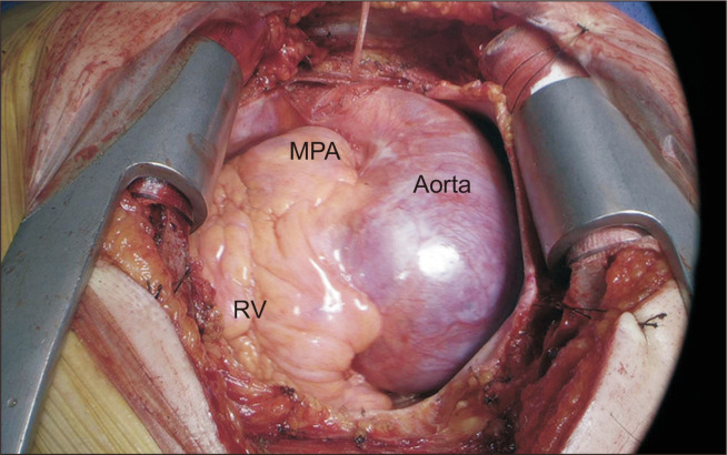
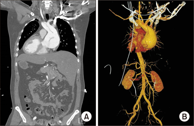
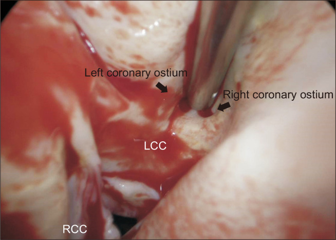
We describe the occurrence of acute type A aortic dissection in a patient with situs inversus totalis. A 37-year-old man presented to the emergency department with acute chest pain. Initial chest X-ray findings showed a right-sided heart and a left-sided liver. Contrast- enhanced computed tomography revealed a Stanford type A acute aortic dissection, aortic root dilatation, and situs inversus totalis. All of the thoracic structures were mirror-image reversed and an abnormal coronary artery was observed. The Bentall operation was performed. This report demonstrates that computed tomography and echocardiography were useful for understanding the anatomy and the presence or absence of concurrent anomalies in a patient with situs inversus totalis. The patient's postoperative course was uneventful.

 求助内容:
求助内容: 应助结果提醒方式:


