Mohammad R Islam, Renhao Luo, Sophia Valaris, Erin B Haley, Hajime Takase, Yinching Iris Chen, Bradford C Dickerson, Karin Schon, Ken Arai, Christopher T Nguyen, Christiane D Wrann
{"title":"扩散张量- mri检测小鼠海马微结构运动诱导的神经可塑性。","authors":"Mohammad R Islam, Renhao Luo, Sophia Valaris, Erin B Haley, Hajime Takase, Yinching Iris Chen, Bradford C Dickerson, Karin Schon, Ken Arai, Christopher T Nguyen, Christiane D Wrann","doi":"10.3233/BPL-190090","DOIUrl":null,"url":null,"abstract":"<p><strong>Background: </strong>Despite considerable research on exercise-induced neuroplasticity in the brain, a major ongoing challenge in translating findings from animal studies to humans is that clinical and preclinical settings employ very different techniques.</p><p><strong>Objective: </strong>Here we aim to bridge this divide by using diffusion tensor imaging MRI (DTI), an advanced imaging technique commonly applied in human studies, in a longitudinal exercise study with mice.</p><p><strong>Methods: </strong>Wild-type mice were exercised using voluntary free-wheel running, and MRI scans were at baseline and after four weeks and nine weeks of running.</p><p><strong>Results: </strong>Both hippocampal volume and fractional anisotropy, a surrogate for microstructural directionality, significantly increased with exercise. In addition, exercise levels correlated with effect size. Histological analysis showed more PDGFR<i>α</i>+ oligodendrocyte precursor cells in the corpus callosum of running mice.</p><p><strong>Conclusions: </strong>These results provide compelling <i>in vivo</i> support for the concept that similar adaptive changes occur in the brains of mice and humans in response to exercise.</p>","PeriodicalId":72451,"journal":{"name":"Brain plasticity (Amsterdam, Netherlands)","volume":"5 2","pages":"147-159"},"PeriodicalIF":0.0000,"publicationDate":"2020-10-01","publicationTypes":"Journal Article","fieldsOfStudy":null,"isOpenAccess":false,"openAccessPdf":"https://sci-hub-pdf.com/10.3233/BPL-190090","citationCount":"9","resultStr":"{\"title\":\"Diffusion tensor-MRI detects exercise-induced neuroplasticity in the hippocampal microstructure in mice.\",\"authors\":\"Mohammad R Islam, Renhao Luo, Sophia Valaris, Erin B Haley, Hajime Takase, Yinching Iris Chen, Bradford C Dickerson, Karin Schon, Ken Arai, Christopher T Nguyen, Christiane D Wrann\",\"doi\":\"10.3233/BPL-190090\",\"DOIUrl\":null,\"url\":null,\"abstract\":\"<p><strong>Background: </strong>Despite considerable research on exercise-induced neuroplasticity in the brain, a major ongoing challenge in translating findings from animal studies to humans is that clinical and preclinical settings employ very different techniques.</p><p><strong>Objective: </strong>Here we aim to bridge this divide by using diffusion tensor imaging MRI (DTI), an advanced imaging technique commonly applied in human studies, in a longitudinal exercise study with mice.</p><p><strong>Methods: </strong>Wild-type mice were exercised using voluntary free-wheel running, and MRI scans were at baseline and after four weeks and nine weeks of running.</p><p><strong>Results: </strong>Both hippocampal volume and fractional anisotropy, a surrogate for microstructural directionality, significantly increased with exercise. In addition, exercise levels correlated with effect size. Histological analysis showed more PDGFR<i>α</i>+ oligodendrocyte precursor cells in the corpus callosum of running mice.</p><p><strong>Conclusions: </strong>These results provide compelling <i>in vivo</i> support for the concept that similar adaptive changes occur in the brains of mice and humans in response to exercise.</p>\",\"PeriodicalId\":72451,\"journal\":{\"name\":\"Brain plasticity (Amsterdam, Netherlands)\",\"volume\":\"5 2\",\"pages\":\"147-159\"},\"PeriodicalIF\":0.0000,\"publicationDate\":\"2020-10-01\",\"publicationTypes\":\"Journal Article\",\"fieldsOfStudy\":null,\"isOpenAccess\":false,\"openAccessPdf\":\"https://sci-hub-pdf.com/10.3233/BPL-190090\",\"citationCount\":\"9\",\"resultStr\":null,\"platform\":\"Semanticscholar\",\"paperid\":null,\"PeriodicalName\":\"Brain plasticity (Amsterdam, Netherlands)\",\"FirstCategoryId\":\"1085\",\"ListUrlMain\":\"https://doi.org/10.3233/BPL-190090\",\"RegionNum\":0,\"RegionCategory\":null,\"ArticlePicture\":[],\"TitleCN\":null,\"AbstractTextCN\":null,\"PMCID\":null,\"EPubDate\":\"\",\"PubModel\":\"\",\"JCR\":\"\",\"JCRName\":\"\",\"Score\":null,\"Total\":0}","platform":"Semanticscholar","paperid":null,"PeriodicalName":"Brain plasticity (Amsterdam, Netherlands)","FirstCategoryId":"1085","ListUrlMain":"https://doi.org/10.3233/BPL-190090","RegionNum":0,"RegionCategory":null,"ArticlePicture":[],"TitleCN":null,"AbstractTextCN":null,"PMCID":null,"EPubDate":"","PubModel":"","JCR":"","JCRName":"","Score":null,"Total":0}
Diffusion tensor-MRI detects exercise-induced neuroplasticity in the hippocampal microstructure in mice.
Background: Despite considerable research on exercise-induced neuroplasticity in the brain, a major ongoing challenge in translating findings from animal studies to humans is that clinical and preclinical settings employ very different techniques.
Objective: Here we aim to bridge this divide by using diffusion tensor imaging MRI (DTI), an advanced imaging technique commonly applied in human studies, in a longitudinal exercise study with mice.
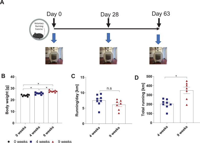
Methods: Wild-type mice were exercised using voluntary free-wheel running, and MRI scans were at baseline and after four weeks and nine weeks of running.
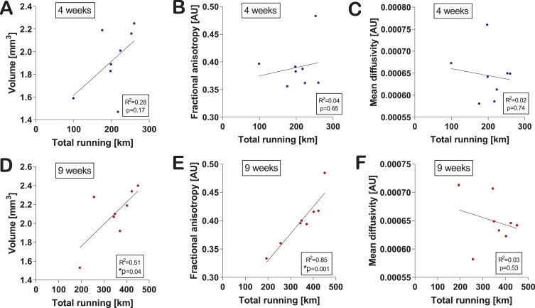
Results: Both hippocampal volume and fractional anisotropy, a surrogate for microstructural directionality, significantly increased with exercise. In addition, exercise levels correlated with effect size. Histological analysis showed more PDGFRα+ oligodendrocyte precursor cells in the corpus callosum of running mice.
Conclusions: These results provide compelling in vivo support for the concept that similar adaptive changes occur in the brains of mice and humans in response to exercise.


 求助内容:
求助内容: 应助结果提醒方式:


