Michael J Colello, Erin R Pichiotino, Stephanie L Tanner, Scott E Porter, Richard W Gurich
{"title":"使用Scout计算机断层扫描(CT)成像预测病理性骨病变。","authors":"Michael J Colello, Erin R Pichiotino, Stephanie L Tanner, Scott E Porter, Richard W Gurich","doi":"10.1155/2020/5105196","DOIUrl":null,"url":null,"abstract":"<p><p>The purpose of this study is to evaluate the benefit of reviewing scout CT images, obtained for routine oncologic surveillance, for the early identification of pathologic bony lesions. A retrospective review was conducted on patients who previously underwent surgical treatment by two orthopedic oncology surgeons at a tertiary care institution from 2009-2019 for pathologic lesions or fractures of the humerus or femur. Radiographic records were reviewed to identify patients in this cohort who had available scout views from CT imaging prior to official diagnosis of the bony lesion or fracture. CT scout images were assessed by two independent reviewers to identify any pathologic lesions, and radiographic reports were reviewed to identify if the lesions were noted by radiology at the time of the initial scan interpretation. One hundred and forty-four patients were identified, and thirty-nine had an available scout CT image prior to official diagnosis of the lesion. Twenty-five patients (64.1%) had lesions identified by authors on scout CT versus only 9 (23.1%) who had lesions that were documented in the initial CT radiologic report. There was a total of 29 lesions identified by the study authors on scout CT, and 19 (65.5%) were not reported in the initial radiographic interpretation with an average interval between observation by authors and official diagnosis of 202 days. Of the impending fractures, three patients (16.7%) went on to complete fracture prior to referral to orthopedics with an average interval between these missed lesions on scout CT and their presentation with fracture of 68 days. This study advocates for the careful review of all scout CT imaging as an essential part of the work up for metastatic disease and encourages all practitioners to utilize this screening tool for the identification of pathologic bony lesions which may help expedite early treatment to reduce patient morbidity.</p>","PeriodicalId":21431,"journal":{"name":"Sarcoma","volume":"2020 ","pages":"5105196"},"PeriodicalIF":0.0000,"publicationDate":"2020-08-06","publicationTypes":"Journal Article","fieldsOfStudy":null,"isOpenAccess":false,"openAccessPdf":"https://sci-hub-pdf.com/10.1155/2020/5105196","citationCount":"1","resultStr":"{\"title\":\"Predicting Pathologic Bone Lesions Using Scout Computed Tomography (CT) Imaging.\",\"authors\":\"Michael J Colello, Erin R Pichiotino, Stephanie L Tanner, Scott E Porter, Richard W Gurich\",\"doi\":\"10.1155/2020/5105196\",\"DOIUrl\":null,\"url\":null,\"abstract\":\"<p><p>The purpose of this study is to evaluate the benefit of reviewing scout CT images, obtained for routine oncologic surveillance, for the early identification of pathologic bony lesions. A retrospective review was conducted on patients who previously underwent surgical treatment by two orthopedic oncology surgeons at a tertiary care institution from 2009-2019 for pathologic lesions or fractures of the humerus or femur. Radiographic records were reviewed to identify patients in this cohort who had available scout views from CT imaging prior to official diagnosis of the bony lesion or fracture. CT scout images were assessed by two independent reviewers to identify any pathologic lesions, and radiographic reports were reviewed to identify if the lesions were noted by radiology at the time of the initial scan interpretation. One hundred and forty-four patients were identified, and thirty-nine had an available scout CT image prior to official diagnosis of the lesion. Twenty-five patients (64.1%) had lesions identified by authors on scout CT versus only 9 (23.1%) who had lesions that were documented in the initial CT radiologic report. There was a total of 29 lesions identified by the study authors on scout CT, and 19 (65.5%) were not reported in the initial radiographic interpretation with an average interval between observation by authors and official diagnosis of 202 days. Of the impending fractures, three patients (16.7%) went on to complete fracture prior to referral to orthopedics with an average interval between these missed lesions on scout CT and their presentation with fracture of 68 days. This study advocates for the careful review of all scout CT imaging as an essential part of the work up for metastatic disease and encourages all practitioners to utilize this screening tool for the identification of pathologic bony lesions which may help expedite early treatment to reduce patient morbidity.</p>\",\"PeriodicalId\":21431,\"journal\":{\"name\":\"Sarcoma\",\"volume\":\"2020 \",\"pages\":\"5105196\"},\"PeriodicalIF\":0.0000,\"publicationDate\":\"2020-08-06\",\"publicationTypes\":\"Journal Article\",\"fieldsOfStudy\":null,\"isOpenAccess\":false,\"openAccessPdf\":\"https://sci-hub-pdf.com/10.1155/2020/5105196\",\"citationCount\":\"1\",\"resultStr\":null,\"platform\":\"Semanticscholar\",\"paperid\":null,\"PeriodicalName\":\"Sarcoma\",\"FirstCategoryId\":\"1085\",\"ListUrlMain\":\"https://doi.org/10.1155/2020/5105196\",\"RegionNum\":0,\"RegionCategory\":null,\"ArticlePicture\":[],\"TitleCN\":null,\"AbstractTextCN\":null,\"PMCID\":null,\"EPubDate\":\"2020/1/1 0:00:00\",\"PubModel\":\"eCollection\",\"JCR\":\"Q2\",\"JCRName\":\"Medicine\",\"Score\":null,\"Total\":0}","platform":"Semanticscholar","paperid":null,"PeriodicalName":"Sarcoma","FirstCategoryId":"1085","ListUrlMain":"https://doi.org/10.1155/2020/5105196","RegionNum":0,"RegionCategory":null,"ArticlePicture":[],"TitleCN":null,"AbstractTextCN":null,"PMCID":null,"EPubDate":"2020/1/1 0:00:00","PubModel":"eCollection","JCR":"Q2","JCRName":"Medicine","Score":null,"Total":0}
Predicting Pathologic Bone Lesions Using Scout Computed Tomography (CT) Imaging.
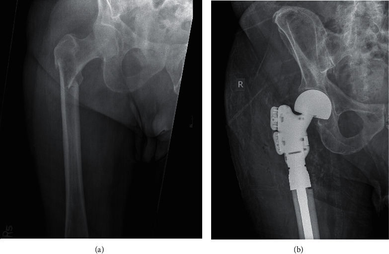
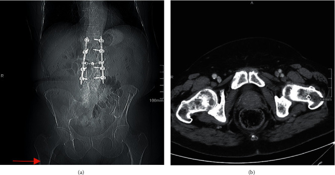
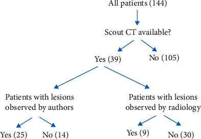
The purpose of this study is to evaluate the benefit of reviewing scout CT images, obtained for routine oncologic surveillance, for the early identification of pathologic bony lesions. A retrospective review was conducted on patients who previously underwent surgical treatment by two orthopedic oncology surgeons at a tertiary care institution from 2009-2019 for pathologic lesions or fractures of the humerus or femur. Radiographic records were reviewed to identify patients in this cohort who had available scout views from CT imaging prior to official diagnosis of the bony lesion or fracture. CT scout images were assessed by two independent reviewers to identify any pathologic lesions, and radiographic reports were reviewed to identify if the lesions were noted by radiology at the time of the initial scan interpretation. One hundred and forty-four patients were identified, and thirty-nine had an available scout CT image prior to official diagnosis of the lesion. Twenty-five patients (64.1%) had lesions identified by authors on scout CT versus only 9 (23.1%) who had lesions that were documented in the initial CT radiologic report. There was a total of 29 lesions identified by the study authors on scout CT, and 19 (65.5%) were not reported in the initial radiographic interpretation with an average interval between observation by authors and official diagnosis of 202 days. Of the impending fractures, three patients (16.7%) went on to complete fracture prior to referral to orthopedics with an average interval between these missed lesions on scout CT and their presentation with fracture of 68 days. This study advocates for the careful review of all scout CT imaging as an essential part of the work up for metastatic disease and encourages all practitioners to utilize this screening tool for the identification of pathologic bony lesions which may help expedite early treatment to reduce patient morbidity.
SarcomaMedicine-Radiology, Nuclear Medicine and Imaging
CiteScore
5.00
自引率
0.00%
发文量
15
审稿时长
14 weeks
期刊介绍:
Sarcoma is dedicated to publishing papers covering all aspects of connective tissue oncology research. It brings together work from scientists and clinicians carrying out a broad range of research in this field, including the basic sciences, molecular biology and pathology and the clinical sciences of epidemiology, surgery, radiotherapy and chemotherapy. High-quality papers concerning the entire range of bone and soft tissue sarcomas in both adults and children, including Kaposi"s sarcoma, are published as well as preclinical and animal studies. This journal provides a central forum for the description of advances in diagnosis, assessment and treatment of this rarely seen, but often mismanaged, group of patients.

 求助内容:
求助内容: 应助结果提醒方式:


