Narong Khuntikeo, Attapol Titapun, Nittaya Chamadol, Wuttisak Boonphongsathien, Prakasit Sa-Ngiamwibool, Simon D Taylor-Robinson, Christopher A Wadsworth, Shuo Zhang, Evdokia M Kardoulaki, Richard R A Syms
{"title":"使用导管线圈对胆管癌进行体外导管内磁共振成像和 T2 映像分析","authors":"Narong Khuntikeo, Attapol Titapun, Nittaya Chamadol, Wuttisak Boonphongsathien, Prakasit Sa-Ngiamwibool, Simon D Taylor-Robinson, Christopher A Wadsworth, Shuo Zhang, Evdokia M Kardoulaki, Richard R A Syms","doi":"10.2147/HMER.S266841","DOIUrl":null,"url":null,"abstract":"<p><strong>Aim: </strong>Diagnostic imaging of early-stage cholangiocarcinoma is challenging. A previous in vitro study of fixed-tissue liver resection specimens investigated T2 mapping as a method of exploiting the locally increased signal-to-noise ratio (SNR) of duodenoscope coils for improved quantitative magnetic resonance imaging (MRI), despite their non-uniform sensitivity. This work applies similar methods to unfixed liver specimens using catheter-based receivers.</p><p><strong>Methods: </strong>Ex vivo intraductal MRI and T2 mapping were carried out at 3T on unfixed resection specimens obtained from cholangiocarcinoma patients immediately after surgery using a catheter coil based on a thin-film magneto-inductive waveguide, inserted directly into an intrahepatic duct.</p><p><strong>Results: </strong>Polypoid intraductal cholangiocarcinoma was imaged using fast spin-echo sequences. High-resolution T2 maps were extracted by fitting of data obtained at different echo times to mono-exponential models, and disease-induced changes were correlated with histopathology. An increase in T2 was found compared with fixed specimens and differences in T2 allowed the resolution of tumour tissue and malignant features such as polypoid morphology.</p><p><strong>Conclusion: </strong>Despite their limited field of view, useful data can be obtained using catheter coils, and T2 mapping offers an effective method of exploiting their local SNR advantage without the need for image correction.</p>","PeriodicalId":12917,"journal":{"name":"Hepatic Medicine : Evidence and Research","volume":"12 ","pages":"107-114"},"PeriodicalIF":1.8000,"publicationDate":"2020-07-27","publicationTypes":"Journal Article","fieldsOfStudy":null,"isOpenAccess":false,"openAccessPdf":"https://www.ncbi.nlm.nih.gov/pmc/articles/PMC7397475/pdf/","citationCount":"0","resultStr":"{\"title\":\"In Vitro Intraductal MRI and T2 Mapping of Cholangiocarcinoma Using Catheter Coils.\",\"authors\":\"Narong Khuntikeo, Attapol Titapun, Nittaya Chamadol, Wuttisak Boonphongsathien, Prakasit Sa-Ngiamwibool, Simon D Taylor-Robinson, Christopher A Wadsworth, Shuo Zhang, Evdokia M Kardoulaki, Richard R A Syms\",\"doi\":\"10.2147/HMER.S266841\",\"DOIUrl\":null,\"url\":null,\"abstract\":\"<p><strong>Aim: </strong>Diagnostic imaging of early-stage cholangiocarcinoma is challenging. A previous in vitro study of fixed-tissue liver resection specimens investigated T2 mapping as a method of exploiting the locally increased signal-to-noise ratio (SNR) of duodenoscope coils for improved quantitative magnetic resonance imaging (MRI), despite their non-uniform sensitivity. This work applies similar methods to unfixed liver specimens using catheter-based receivers.</p><p><strong>Methods: </strong>Ex vivo intraductal MRI and T2 mapping were carried out at 3T on unfixed resection specimens obtained from cholangiocarcinoma patients immediately after surgery using a catheter coil based on a thin-film magneto-inductive waveguide, inserted directly into an intrahepatic duct.</p><p><strong>Results: </strong>Polypoid intraductal cholangiocarcinoma was imaged using fast spin-echo sequences. High-resolution T2 maps were extracted by fitting of data obtained at different echo times to mono-exponential models, and disease-induced changes were correlated with histopathology. An increase in T2 was found compared with fixed specimens and differences in T2 allowed the resolution of tumour tissue and malignant features such as polypoid morphology.</p><p><strong>Conclusion: </strong>Despite their limited field of view, useful data can be obtained using catheter coils, and T2 mapping offers an effective method of exploiting their local SNR advantage without the need for image correction.</p>\",\"PeriodicalId\":12917,\"journal\":{\"name\":\"Hepatic Medicine : Evidence and Research\",\"volume\":\"12 \",\"pages\":\"107-114\"},\"PeriodicalIF\":1.8000,\"publicationDate\":\"2020-07-27\",\"publicationTypes\":\"Journal Article\",\"fieldsOfStudy\":null,\"isOpenAccess\":false,\"openAccessPdf\":\"https://www.ncbi.nlm.nih.gov/pmc/articles/PMC7397475/pdf/\",\"citationCount\":\"0\",\"resultStr\":null,\"platform\":\"Semanticscholar\",\"paperid\":null,\"PeriodicalName\":\"Hepatic Medicine : Evidence and Research\",\"FirstCategoryId\":\"1085\",\"ListUrlMain\":\"https://doi.org/10.2147/HMER.S266841\",\"RegionNum\":0,\"RegionCategory\":null,\"ArticlePicture\":[],\"TitleCN\":null,\"AbstractTextCN\":null,\"PMCID\":null,\"EPubDate\":\"2020/1/1 0:00:00\",\"PubModel\":\"eCollection\",\"JCR\":\"Q2\",\"JCRName\":\"GASTROENTEROLOGY & HEPATOLOGY\",\"Score\":null,\"Total\":0}","platform":"Semanticscholar","paperid":null,"PeriodicalName":"Hepatic Medicine : Evidence and Research","FirstCategoryId":"1085","ListUrlMain":"https://doi.org/10.2147/HMER.S266841","RegionNum":0,"RegionCategory":null,"ArticlePicture":[],"TitleCN":null,"AbstractTextCN":null,"PMCID":null,"EPubDate":"2020/1/1 0:00:00","PubModel":"eCollection","JCR":"Q2","JCRName":"GASTROENTEROLOGY & HEPATOLOGY","Score":null,"Total":0}
In Vitro Intraductal MRI and T2 Mapping of Cholangiocarcinoma Using Catheter Coils.
Aim: Diagnostic imaging of early-stage cholangiocarcinoma is challenging. A previous in vitro study of fixed-tissue liver resection specimens investigated T2 mapping as a method of exploiting the locally increased signal-to-noise ratio (SNR) of duodenoscope coils for improved quantitative magnetic resonance imaging (MRI), despite their non-uniform sensitivity. This work applies similar methods to unfixed liver specimens using catheter-based receivers.
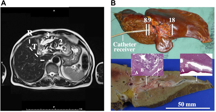
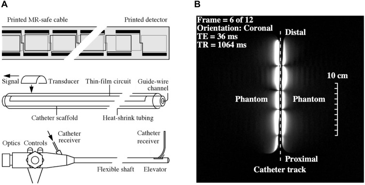
Methods: Ex vivo intraductal MRI and T2 mapping were carried out at 3T on unfixed resection specimens obtained from cholangiocarcinoma patients immediately after surgery using a catheter coil based on a thin-film magneto-inductive waveguide, inserted directly into an intrahepatic duct.
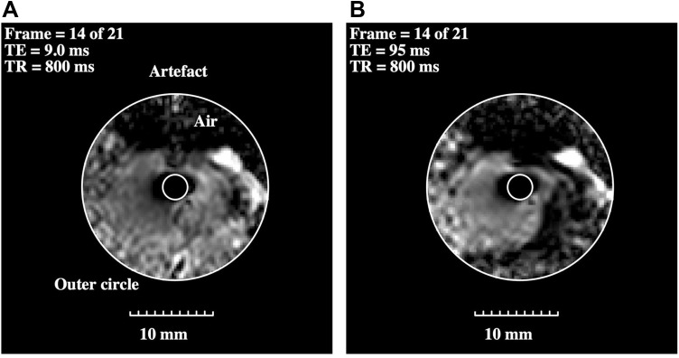
Results: Polypoid intraductal cholangiocarcinoma was imaged using fast spin-echo sequences. High-resolution T2 maps were extracted by fitting of data obtained at different echo times to mono-exponential models, and disease-induced changes were correlated with histopathology. An increase in T2 was found compared with fixed specimens and differences in T2 allowed the resolution of tumour tissue and malignant features such as polypoid morphology.
Conclusion: Despite their limited field of view, useful data can be obtained using catheter coils, and T2 mapping offers an effective method of exploiting their local SNR advantage without the need for image correction.
期刊介绍:
Hepatic Medicine: Evidence and Research is an international, peer-reviewed, open access, online journal. Publishing original research, reports, editorials, reviews and commentaries on all aspects of adult and pediatric hepatology in the clinic and laboratory including the following topics: Pathology, pathophysiology of hepatic disease Investigation and treatment of hepatic disease Pharmacology of drugs used for the treatment of hepatic disease Although the main focus of the journal is to publish research and clinical results in humans; preclinical, animal and in vitro studies will be published where they will shed light on disease processes and potential new therapies. Issues of patient safety and quality of care will also be considered. As of 1st April 2019, Hepatic Medicine: Evidence and Research will no longer consider meta-analyses for publication.

 求助内容:
求助内容: 应助结果提醒方式:


