{"title":"从磷酸酶角度看 G2 到 M 的转变:减数分裂的新视野。","authors":"Tom Lemonnier, Aude Dupré, Catherine Jessus","doi":"10.1186/s13008-020-00065-2","DOIUrl":null,"url":null,"abstract":"<p><p>Cell division is orchestrated by the phosphorylation and dephosphorylation of thousands of proteins. These post-translational modifications underlie the molecular cascades converging to the activation of the universal mitotic kinase, Cdk1, and entry into cell division. They also govern the structural events that sustain the mechanics of cell division. While the role of protein kinases in mitosis has been well documented by decades of investigations, little was known regarding the control of protein phosphatases until the recent years. However, the regulation of phosphatase activities is as essential as kinases in controlling the activation of Cdk1 to enter M-phase. The regulation and the function of phosphatases result from post-translational modifications but also from the combinatorial association between conserved catalytic subunits and regulatory subunits that drive their substrate specificity, their cellular localization and their activity. It now appears that sequential dephosphorylations orchestrated by a network of phosphatase activities trigger Cdk1 activation and then order the structural events necessary for the timely execution of cell division. This review discusses a series of recent works describing the important roles played by protein phosphatases for the proper regulation of meiotic division. Many breakthroughs in the field of cell cycle research came from studies on oocyte meiotic divisions. Indeed, the meiotic division shares most of the molecular regulators with mitosis. The natural arrests of oocytes in G2 and in M-phase, the giant size of these cells, the variety of model species allowing either biochemical or imaging as well as genetics approaches explain why the process of meiosis has served as an historical model to decipher signalling pathways involved in the G2-to-M transition. The review especially highlights how the phosphatase PP2A-B55δ critically orchestrates the timing of meiosis resumption in amphibian oocytes. By opposing the kinase PKA, PP2A-B55δ controls the release of the G2 arrest through the dephosphorylation of their substrate, Arpp19. Few hours later, the inhibition of PP2A-B55δ by Arpp19 releases its opposing kinase, Cdk1, and triggers M-phase. In coordination with a variety of phosphatases and kinases, the PP2A-B55δ/Arpp19 duo therefore emerges as the key effector of the G2-to-M transition.</p>","PeriodicalId":49263,"journal":{"name":"Cell Division","volume":"15 ","pages":"9"},"PeriodicalIF":2.2000,"publicationDate":"2020-05-25","publicationTypes":"Journal Article","fieldsOfStudy":null,"isOpenAccess":false,"openAccessPdf":"https://www.ncbi.nlm.nih.gov/pmc/articles/PMC7249327/pdf/","citationCount":"0","resultStr":"{\"title\":\"The G2-to-M transition from a phosphatase perspective: a new vision of the meiotic division.\",\"authors\":\"Tom Lemonnier, Aude Dupré, Catherine Jessus\",\"doi\":\"10.1186/s13008-020-00065-2\",\"DOIUrl\":null,\"url\":null,\"abstract\":\"<p><p>Cell division is orchestrated by the phosphorylation and dephosphorylation of thousands of proteins. These post-translational modifications underlie the molecular cascades converging to the activation of the universal mitotic kinase, Cdk1, and entry into cell division. They also govern the structural events that sustain the mechanics of cell division. While the role of protein kinases in mitosis has been well documented by decades of investigations, little was known regarding the control of protein phosphatases until the recent years. However, the regulation of phosphatase activities is as essential as kinases in controlling the activation of Cdk1 to enter M-phase. The regulation and the function of phosphatases result from post-translational modifications but also from the combinatorial association between conserved catalytic subunits and regulatory subunits that drive their substrate specificity, their cellular localization and their activity. It now appears that sequential dephosphorylations orchestrated by a network of phosphatase activities trigger Cdk1 activation and then order the structural events necessary for the timely execution of cell division. This review discusses a series of recent works describing the important roles played by protein phosphatases for the proper regulation of meiotic division. Many breakthroughs in the field of cell cycle research came from studies on oocyte meiotic divisions. Indeed, the meiotic division shares most of the molecular regulators with mitosis. The natural arrests of oocytes in G2 and in M-phase, the giant size of these cells, the variety of model species allowing either biochemical or imaging as well as genetics approaches explain why the process of meiosis has served as an historical model to decipher signalling pathways involved in the G2-to-M transition. The review especially highlights how the phosphatase PP2A-B55δ critically orchestrates the timing of meiosis resumption in amphibian oocytes. By opposing the kinase PKA, PP2A-B55δ controls the release of the G2 arrest through the dephosphorylation of their substrate, Arpp19. Few hours later, the inhibition of PP2A-B55δ by Arpp19 releases its opposing kinase, Cdk1, and triggers M-phase. In coordination with a variety of phosphatases and kinases, the PP2A-B55δ/Arpp19 duo therefore emerges as the key effector of the G2-to-M transition.</p>\",\"PeriodicalId\":49263,\"journal\":{\"name\":\"Cell Division\",\"volume\":\"15 \",\"pages\":\"9\"},\"PeriodicalIF\":2.2000,\"publicationDate\":\"2020-05-25\",\"publicationTypes\":\"Journal Article\",\"fieldsOfStudy\":null,\"isOpenAccess\":false,\"openAccessPdf\":\"https://www.ncbi.nlm.nih.gov/pmc/articles/PMC7249327/pdf/\",\"citationCount\":\"0\",\"resultStr\":null,\"platform\":\"Semanticscholar\",\"paperid\":null,\"PeriodicalName\":\"Cell Division\",\"FirstCategoryId\":\"99\",\"ListUrlMain\":\"https://doi.org/10.1186/s13008-020-00065-2\",\"RegionNum\":4,\"RegionCategory\":\"生物学\",\"ArticlePicture\":[],\"TitleCN\":null,\"AbstractTextCN\":null,\"PMCID\":null,\"EPubDate\":\"2020/1/1 0:00:00\",\"PubModel\":\"eCollection\",\"JCR\":\"Q3\",\"JCRName\":\"CELL BIOLOGY\",\"Score\":null,\"Total\":0}","platform":"Semanticscholar","paperid":null,"PeriodicalName":"Cell Division","FirstCategoryId":"99","ListUrlMain":"https://doi.org/10.1186/s13008-020-00065-2","RegionNum":4,"RegionCategory":"生物学","ArticlePicture":[],"TitleCN":null,"AbstractTextCN":null,"PMCID":null,"EPubDate":"2020/1/1 0:00:00","PubModel":"eCollection","JCR":"Q3","JCRName":"CELL BIOLOGY","Score":null,"Total":0}
引用次数: 0
摘要
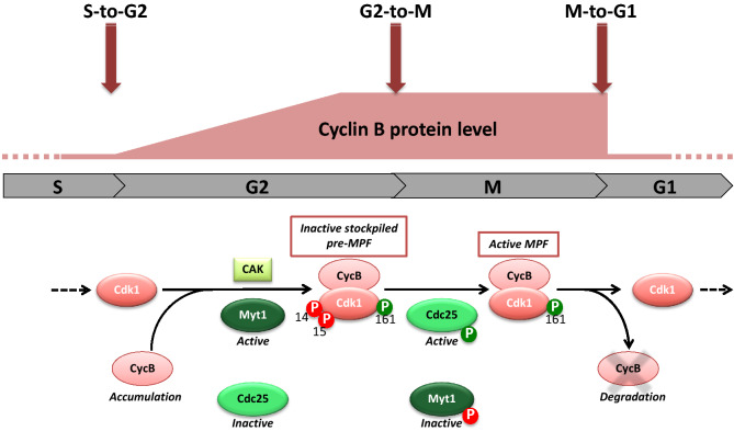
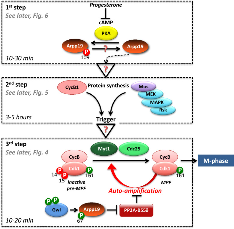
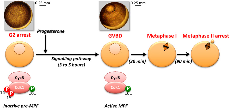
细胞分裂是由数千种蛋白质的磷酸化和去磷酸化协调进行的。这些翻译后修饰是激活有丝分裂通用激酶 Cdk1 和进入细胞分裂的分子级联的基础。它们还控制着维持细胞分裂机制的结构事件。几十年来,蛋白激酶在有丝分裂中的作用已被大量研究证实,但直到最近几年,人们才对蛋白磷酸酶的调控作用知之甚少。然而,在控制 Cdk1 进入 M 期的过程中,磷酸酶活性的调节与激酶一样重要。磷酸酶的调控和功能来自翻译后修饰,也来自保守的催化亚基和调控亚基之间的组合关联,这些关联驱动着磷酸酶的底物特异性、细胞定位和活性。现在看来,由磷酸酶活动网络协调的连续去磷酸化会触发 Cdk1 激活,然后对及时执行细胞分裂所需的结构事件进行排序。这篇综述讨论了最近一系列描述蛋白磷酸酶在正确调控减数分裂方面所发挥的重要作用的研究成果。细胞周期研究领域的许多突破都来自对卵母细胞减数分裂的研究。事实上,减数分裂与有丝分裂有着相同的分子调节机制。卵母细胞在 G2 期和 M 期的自然停滞、这些细胞的巨大体积、允许采用生化或成像以及遗传学方法的各种模型物种,都解释了为什么减数分裂过程一直是破译 G2 到 M 转变过程中信号通路的历史性模型。这篇综述特别强调了磷酸酶 PP2A-B55δ 如何对两栖动物卵母细胞减数分裂恢复的时间进行关键性的协调。通过对抗激酶 PKA,PP2A-B55δ 通过其底物 Arpp19 的去磷酸化控制 G2 停顿的释放。几小时后,Arpp19 对 PP2A-B55δ 的抑制释放了其对立激酶 Cdk1,并引发 M 期。因此,PP2A-B55δ/Arpp19 二人组合与多种磷酸酶和激酶配合,成为 G2 向 M 过渡的关键效应器。
The G2-to-M transition from a phosphatase perspective: a new vision of the meiotic division.
Cell division is orchestrated by the phosphorylation and dephosphorylation of thousands of proteins. These post-translational modifications underlie the molecular cascades converging to the activation of the universal mitotic kinase, Cdk1, and entry into cell division. They also govern the structural events that sustain the mechanics of cell division. While the role of protein kinases in mitosis has been well documented by decades of investigations, little was known regarding the control of protein phosphatases until the recent years. However, the regulation of phosphatase activities is as essential as kinases in controlling the activation of Cdk1 to enter M-phase. The regulation and the function of phosphatases result from post-translational modifications but also from the combinatorial association between conserved catalytic subunits and regulatory subunits that drive their substrate specificity, their cellular localization and their activity. It now appears that sequential dephosphorylations orchestrated by a network of phosphatase activities trigger Cdk1 activation and then order the structural events necessary for the timely execution of cell division. This review discusses a series of recent works describing the important roles played by protein phosphatases for the proper regulation of meiotic division. Many breakthroughs in the field of cell cycle research came from studies on oocyte meiotic divisions. Indeed, the meiotic division shares most of the molecular regulators with mitosis. The natural arrests of oocytes in G2 and in M-phase, the giant size of these cells, the variety of model species allowing either biochemical or imaging as well as genetics approaches explain why the process of meiosis has served as an historical model to decipher signalling pathways involved in the G2-to-M transition. The review especially highlights how the phosphatase PP2A-B55δ critically orchestrates the timing of meiosis resumption in amphibian oocytes. By opposing the kinase PKA, PP2A-B55δ controls the release of the G2 arrest through the dephosphorylation of their substrate, Arpp19. Few hours later, the inhibition of PP2A-B55δ by Arpp19 releases its opposing kinase, Cdk1, and triggers M-phase. In coordination with a variety of phosphatases and kinases, the PP2A-B55δ/Arpp19 duo therefore emerges as the key effector of the G2-to-M transition.
期刊介绍:
Cell Division is an open access, peer-reviewed journal that encompasses all the molecular aspects of cell cycle control and cancer, cell growth, proliferation, survival, differentiation, signalling, gene transcription, protein synthesis, genome integrity, chromosome stability, centrosome duplication, DNA damage and DNA repair.
Cell Division provides an online forum for the cell-cycle community that aims to publish articles on all exciting aspects of cell-cycle research and to bridge the gap between models of cell cycle regulation, development, and cancer biology. This forum is driven by specialized and timely research articles, reviews and commentaries focused on this fast moving field, providing an invaluable tool for cell-cycle biologists.
Cell Division publishes articles in areas which includes, but not limited to:
DNA replication, cell fate decisions, cell cycle & development
Cell proliferation, mitosis, spindle assembly checkpoint, ubiquitin mediated degradation
DNA damage & repair
Apoptosis & cell death
 求助内容:
求助内容: 应助结果提醒方式:


