Osama A Sorour, Nihaal Mehta, Caroline R Baumal, Akihiro Ishibazawa, Keke Liu, Eleni K Konstantinou, Sarah Martin, Phillip Braun, A Yasin Alibhai, Malvika Arya, Andre J Witkin, Jay S Duker, Nadia K Waheed
{"title":"光学相干断层血管造影显示抗vegf治疗后视网膜微血管异常的形态学改变。","authors":"Osama A Sorour, Nihaal Mehta, Caroline R Baumal, Akihiro Ishibazawa, Keke Liu, Eleni K Konstantinou, Sarah Martin, Phillip Braun, A Yasin Alibhai, Malvika Arya, Andre J Witkin, Jay S Duker, Nadia K Waheed","doi":"10.1186/s40662-020-00195-2","DOIUrl":null,"url":null,"abstract":"<p><strong>Background: </strong>To examine the baseline morphological characteristics and alterations in intraretinal microvascular abnormalities (IRMAs) in response to anti-vascular endothelial growth factor (anti-VEGF) treatment, documented by optical coherence tomography angiography (OCTA) in diabetic eyes.</p><p><strong>Methods: </strong>In this retrospective study, IRMAs were evaluated with multimodal imaging (fundus photography, fluorescein angiography, OCTA) in treatment-naïve diabetic eyes before and after anti-VEGF treatment for diabetic macular edema (DME) and/or proliferative diabetic retinopathy (PDR) and compared to diabetic control eyes with similar diabetic retinopathy (DR) severity that did not receive anti-VEGF therapy. The morphological characteristics of IRMAs on enface OCTA imaging were graded by masked readers at baseline, then after anti-VEGF therapy in treated eyes or after observation in control eyes. Characterization of interval changes in an IRMA were based on the following parameters: branching, vessel caliber and area of adjacent capillary non-perfusion.</p><p><strong>Results: </strong>The treated group included 45 IRMA foci from 15 eyes of 11 patients, while the control group included 27 IRMA foci from 15 eyes of 14 patients. Following anti-VEGF treatment, enface OCTA demonstrated that 14 foci of IRMA (31%) demonstrated regression with normalization of appearance of the capillary bed, 20 IRMAs (44%) remained unchanged, six IRMAs (13%) progressed with enlargement or development of new IRMAs and five IRMAs (11%) demonstrated complete obliteration defined as IRMA disappearance with advancing capillary drop-out. In the control group, 17 IRMA (63%) remained stable, 8 IRMAs (29.6%) progressed and 2 experienced total obliteration (7.4%). The difference in rank order between the two groups was statistically significant (<i>p</i> = 0.022).</p><p><strong>Conclusions: </strong>In eyes with DR status post anti-VEGF therapy, foci of IRMAs have a variable course demonstrating one of four possible outcomes: regression, stability, progression or complete obliteration. In contrast, none of the untreated control diabetic eyes demonstrated regression of IRMAs, consistent with known progression of DR severity in high risk eyes. Morphologic evaluation of IRMAs with OCTA may help to monitor changes in retinal blood flow as well as the response to anti-VEGF treatment.</p>","PeriodicalId":520624,"journal":{"name":"Eye and vision (London, England)","volume":" ","pages":"29"},"PeriodicalIF":0.0000,"publicationDate":"2020-06-01","publicationTypes":"Journal Article","fieldsOfStudy":null,"isOpenAccess":false,"openAccessPdf":"https://sci-hub-pdf.com/10.1186/s40662-020-00195-2","citationCount":"8","resultStr":"{\"title\":\"Morphological changes in intraretinal microvascular abnormalities after anti-VEGF therapy visualized on optical coherence tomography angiography.\",\"authors\":\"Osama A Sorour, Nihaal Mehta, Caroline R Baumal, Akihiro Ishibazawa, Keke Liu, Eleni K Konstantinou, Sarah Martin, Phillip Braun, A Yasin Alibhai, Malvika Arya, Andre J Witkin, Jay S Duker, Nadia K Waheed\",\"doi\":\"10.1186/s40662-020-00195-2\",\"DOIUrl\":null,\"url\":null,\"abstract\":\"<p><strong>Background: </strong>To examine the baseline morphological characteristics and alterations in intraretinal microvascular abnormalities (IRMAs) in response to anti-vascular endothelial growth factor (anti-VEGF) treatment, documented by optical coherence tomography angiography (OCTA) in diabetic eyes.</p><p><strong>Methods: </strong>In this retrospective study, IRMAs were evaluated with multimodal imaging (fundus photography, fluorescein angiography, OCTA) in treatment-naïve diabetic eyes before and after anti-VEGF treatment for diabetic macular edema (DME) and/or proliferative diabetic retinopathy (PDR) and compared to diabetic control eyes with similar diabetic retinopathy (DR) severity that did not receive anti-VEGF therapy. The morphological characteristics of IRMAs on enface OCTA imaging were graded by masked readers at baseline, then after anti-VEGF therapy in treated eyes or after observation in control eyes. Characterization of interval changes in an IRMA were based on the following parameters: branching, vessel caliber and area of adjacent capillary non-perfusion.</p><p><strong>Results: </strong>The treated group included 45 IRMA foci from 15 eyes of 11 patients, while the control group included 27 IRMA foci from 15 eyes of 14 patients. Following anti-VEGF treatment, enface OCTA demonstrated that 14 foci of IRMA (31%) demonstrated regression with normalization of appearance of the capillary bed, 20 IRMAs (44%) remained unchanged, six IRMAs (13%) progressed with enlargement or development of new IRMAs and five IRMAs (11%) demonstrated complete obliteration defined as IRMA disappearance with advancing capillary drop-out. In the control group, 17 IRMA (63%) remained stable, 8 IRMAs (29.6%) progressed and 2 experienced total obliteration (7.4%). The difference in rank order between the two groups was statistically significant (<i>p</i> = 0.022).</p><p><strong>Conclusions: </strong>In eyes with DR status post anti-VEGF therapy, foci of IRMAs have a variable course demonstrating one of four possible outcomes: regression, stability, progression or complete obliteration. In contrast, none of the untreated control diabetic eyes demonstrated regression of IRMAs, consistent with known progression of DR severity in high risk eyes. Morphologic evaluation of IRMAs with OCTA may help to monitor changes in retinal blood flow as well as the response to anti-VEGF treatment.</p>\",\"PeriodicalId\":520624,\"journal\":{\"name\":\"Eye and vision (London, England)\",\"volume\":\" \",\"pages\":\"29\"},\"PeriodicalIF\":0.0000,\"publicationDate\":\"2020-06-01\",\"publicationTypes\":\"Journal Article\",\"fieldsOfStudy\":null,\"isOpenAccess\":false,\"openAccessPdf\":\"https://sci-hub-pdf.com/10.1186/s40662-020-00195-2\",\"citationCount\":\"8\",\"resultStr\":null,\"platform\":\"Semanticscholar\",\"paperid\":null,\"PeriodicalName\":\"Eye and vision (London, England)\",\"FirstCategoryId\":\"3\",\"ListUrlMain\":\"https://doi.org/10.1186/s40662-020-00195-2\",\"RegionNum\":0,\"RegionCategory\":null,\"ArticlePicture\":[],\"TitleCN\":null,\"AbstractTextCN\":null,\"PMCID\":null,\"EPubDate\":\"2020/1/1 0:00:00\",\"PubModel\":\"eCollection\",\"JCR\":\"\",\"JCRName\":\"\",\"Score\":null,\"Total\":0}","platform":"Semanticscholar","paperid":null,"PeriodicalName":"Eye and vision (London, England)","FirstCategoryId":"3","ListUrlMain":"https://doi.org/10.1186/s40662-020-00195-2","RegionNum":0,"RegionCategory":null,"ArticlePicture":[],"TitleCN":null,"AbstractTextCN":null,"PMCID":null,"EPubDate":"2020/1/1 0:00:00","PubModel":"eCollection","JCR":"","JCRName":"","Score":null,"Total":0}
Morphological changes in intraretinal microvascular abnormalities after anti-VEGF therapy visualized on optical coherence tomography angiography.
Background: To examine the baseline morphological characteristics and alterations in intraretinal microvascular abnormalities (IRMAs) in response to anti-vascular endothelial growth factor (anti-VEGF) treatment, documented by optical coherence tomography angiography (OCTA) in diabetic eyes.
Methods: In this retrospective study, IRMAs were evaluated with multimodal imaging (fundus photography, fluorescein angiography, OCTA) in treatment-naïve diabetic eyes before and after anti-VEGF treatment for diabetic macular edema (DME) and/or proliferative diabetic retinopathy (PDR) and compared to diabetic control eyes with similar diabetic retinopathy (DR) severity that did not receive anti-VEGF therapy. The morphological characteristics of IRMAs on enface OCTA imaging were graded by masked readers at baseline, then after anti-VEGF therapy in treated eyes or after observation in control eyes. Characterization of interval changes in an IRMA were based on the following parameters: branching, vessel caliber and area of adjacent capillary non-perfusion.
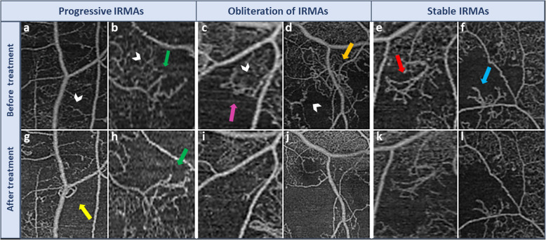
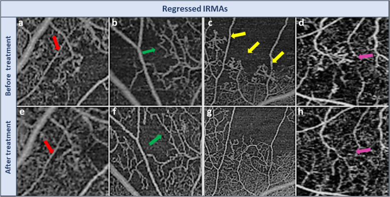
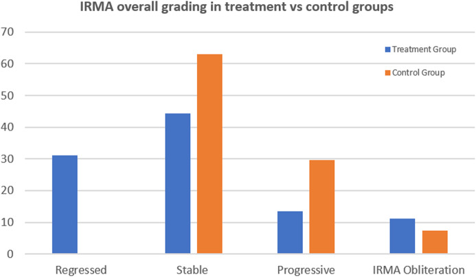
Results: The treated group included 45 IRMA foci from 15 eyes of 11 patients, while the control group included 27 IRMA foci from 15 eyes of 14 patients. Following anti-VEGF treatment, enface OCTA demonstrated that 14 foci of IRMA (31%) demonstrated regression with normalization of appearance of the capillary bed, 20 IRMAs (44%) remained unchanged, six IRMAs (13%) progressed with enlargement or development of new IRMAs and five IRMAs (11%) demonstrated complete obliteration defined as IRMA disappearance with advancing capillary drop-out. In the control group, 17 IRMA (63%) remained stable, 8 IRMAs (29.6%) progressed and 2 experienced total obliteration (7.4%). The difference in rank order between the two groups was statistically significant (p = 0.022).
Conclusions: In eyes with DR status post anti-VEGF therapy, foci of IRMAs have a variable course demonstrating one of four possible outcomes: regression, stability, progression or complete obliteration. In contrast, none of the untreated control diabetic eyes demonstrated regression of IRMAs, consistent with known progression of DR severity in high risk eyes. Morphologic evaluation of IRMAs with OCTA may help to monitor changes in retinal blood flow as well as the response to anti-VEGF treatment.

 求助内容:
求助内容: 应助结果提醒方式:


