{"title":"包封的鼻窦神经鞘瘤模拟鼻鼻息肉。","authors":"Abdulrazak Ajiya, Nafisatu Bello-Muhammad","doi":"10.4103/nmj.NMJ_104_19","DOIUrl":null,"url":null,"abstract":"<p><p>Extracranial schwannomas are uncommon neoplasms of the sinonasal tract arising from peripheral nerve shealth. Mostly acapsulated on histology, but few cases of encapsulated schwannomas have been reported. Its symptoms are nonspecific and initial clinical diagnosis is frequently missed. We report a 13-year-old boy with a huge, encapsulated sinonasal schwannoma initially thought to be an antrochoanal polyp. Computed tomography scan demonstrated a huge irregularly shaped mildly enhancing isodense mass in the right nasal cavity with lateral extension to the ipsilateral maxillary sinus, superior extension into the ethmoids and frontal sinuses and posteriorly into the nasopharynx. The tumour was completely excised via a lateral rhinotomy and patient is still on follow-up.</p>","PeriodicalId":19223,"journal":{"name":"Nigerian Medical Journal : Journal of the Nigeria Medical Association","volume":"60 6","pages":"326-329"},"PeriodicalIF":0.0000,"publicationDate":"2019-11-01","publicationTypes":"Journal Article","fieldsOfStudy":null,"isOpenAccess":false,"openAccessPdf":"https://ftp.ncbi.nlm.nih.gov/pub/pmc/oa_pdf/9d/2a/NMJ-60-326.PMC7053271.pdf","citationCount":"1","resultStr":"{\"title\":\"Encapsulated Sinonasal Schwannoma Mimicking an Antrochoanal Polyp.\",\"authors\":\"Abdulrazak Ajiya, Nafisatu Bello-Muhammad\",\"doi\":\"10.4103/nmj.NMJ_104_19\",\"DOIUrl\":null,\"url\":null,\"abstract\":\"<p><p>Extracranial schwannomas are uncommon neoplasms of the sinonasal tract arising from peripheral nerve shealth. Mostly acapsulated on histology, but few cases of encapsulated schwannomas have been reported. Its symptoms are nonspecific and initial clinical diagnosis is frequently missed. We report a 13-year-old boy with a huge, encapsulated sinonasal schwannoma initially thought to be an antrochoanal polyp. Computed tomography scan demonstrated a huge irregularly shaped mildly enhancing isodense mass in the right nasal cavity with lateral extension to the ipsilateral maxillary sinus, superior extension into the ethmoids and frontal sinuses and posteriorly into the nasopharynx. The tumour was completely excised via a lateral rhinotomy and patient is still on follow-up.</p>\",\"PeriodicalId\":19223,\"journal\":{\"name\":\"Nigerian Medical Journal : Journal of the Nigeria Medical Association\",\"volume\":\"60 6\",\"pages\":\"326-329\"},\"PeriodicalIF\":0.0000,\"publicationDate\":\"2019-11-01\",\"publicationTypes\":\"Journal Article\",\"fieldsOfStudy\":null,\"isOpenAccess\":false,\"openAccessPdf\":\"https://ftp.ncbi.nlm.nih.gov/pub/pmc/oa_pdf/9d/2a/NMJ-60-326.PMC7053271.pdf\",\"citationCount\":\"1\",\"resultStr\":null,\"platform\":\"Semanticscholar\",\"paperid\":null,\"PeriodicalName\":\"Nigerian Medical Journal : Journal of the Nigeria Medical Association\",\"FirstCategoryId\":\"1085\",\"ListUrlMain\":\"https://doi.org/10.4103/nmj.NMJ_104_19\",\"RegionNum\":0,\"RegionCategory\":null,\"ArticlePicture\":[],\"TitleCN\":null,\"AbstractTextCN\":null,\"PMCID\":null,\"EPubDate\":\"2020/2/24 0:00:00\",\"PubModel\":\"Epub\",\"JCR\":\"\",\"JCRName\":\"\",\"Score\":null,\"Total\":0}","platform":"Semanticscholar","paperid":null,"PeriodicalName":"Nigerian Medical Journal : Journal of the Nigeria Medical Association","FirstCategoryId":"1085","ListUrlMain":"https://doi.org/10.4103/nmj.NMJ_104_19","RegionNum":0,"RegionCategory":null,"ArticlePicture":[],"TitleCN":null,"AbstractTextCN":null,"PMCID":null,"EPubDate":"2020/2/24 0:00:00","PubModel":"Epub","JCR":"","JCRName":"","Score":null,"Total":0}
Encapsulated Sinonasal Schwannoma Mimicking an Antrochoanal Polyp.
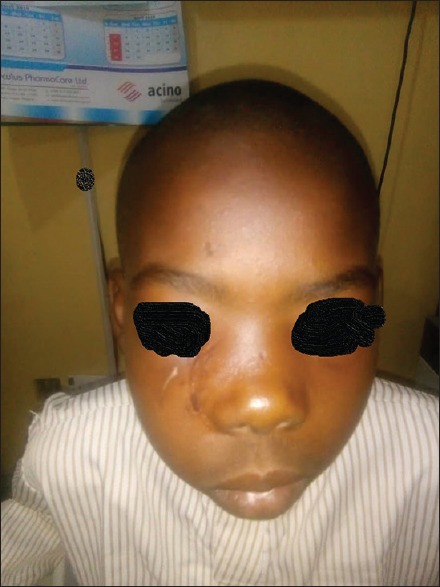
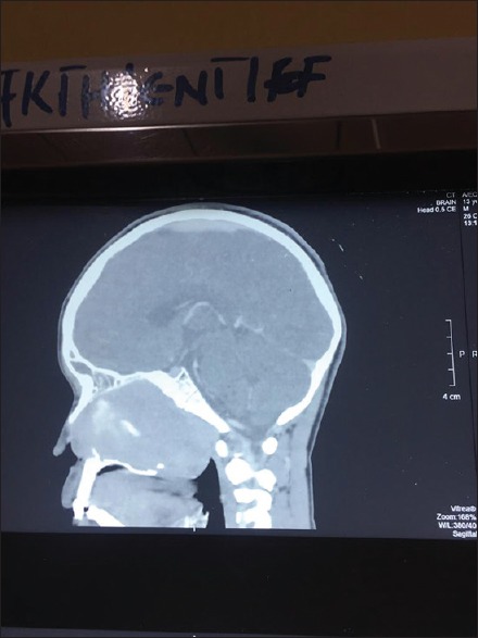
Extracranial schwannomas are uncommon neoplasms of the sinonasal tract arising from peripheral nerve shealth. Mostly acapsulated on histology, but few cases of encapsulated schwannomas have been reported. Its symptoms are nonspecific and initial clinical diagnosis is frequently missed. We report a 13-year-old boy with a huge, encapsulated sinonasal schwannoma initially thought to be an antrochoanal polyp. Computed tomography scan demonstrated a huge irregularly shaped mildly enhancing isodense mass in the right nasal cavity with lateral extension to the ipsilateral maxillary sinus, superior extension into the ethmoids and frontal sinuses and posteriorly into the nasopharynx. The tumour was completely excised via a lateral rhinotomy and patient is still on follow-up.


 求助内容:
求助内容: 应助结果提醒方式:


