Stefano Baiocchi, Cosimo Mazzotta, Arianna Sgheri, Alessandro Di Maggio, Simone Alex Bagaglia, Matteo Posarelli, Leonardo Ciompi, Alessandro Meduri, Gian Marco Tosi
{"title":"体内共聚焦显微镜:对开角型青光眼患者行XEN-Gel植入、小梁切除术或药物治疗后结膜和角膜表面的定性研究。","authors":"Stefano Baiocchi, Cosimo Mazzotta, Arianna Sgheri, Alessandro Di Maggio, Simone Alex Bagaglia, Matteo Posarelli, Leonardo Ciompi, Alessandro Meduri, Gian Marco Tosi","doi":"10.1186/s40662-020-00181-8","DOIUrl":null,"url":null,"abstract":"<p><strong>Purpose: </strong>Assessing the quality of the ocular surface by in vivo scanning laser confocal microscopy (IVCM) in primary open angle glaucoma (POAG) patients treated by Xen 45 Gel Stent, medical therapy and trabeculectomy.</p><p><strong>Methods: </strong>Retrospective, single-center, single-masked, comparative study including 60 eyes of 30 patients (mean age 61.16 ± 10 years) affected by POAG. Eyes were divided into 3 groups: Group 1 eyes underwent the Xen 45 Gel Stent procedure, Group 2 eyes were under medical therapy, Group 3 eyes were surgically treated by trabeculectomy. All patients underwent HRT II IVCM analysis of cornea, limbus, conjunctiva, sub-tenionian space and sclera.</p><p><strong>Results: </strong>The Xen 45 Gel stent, if properly positioned in the sub-conjunctival space preserves goblet cells and limits ocular surface inflammation. Regular corneal epithelial cells with micro-cysts, and normo-reflective sub-epithelial nerve plexus are documented by IVCM. In sub Tenon's implants an alternative lamellar intra-scleral filtration is detectable. Combined surgical procedures show a noticeable number of inflammatory cells with rare micro-cysts. Post-trabeculectomy inflammatory reaction is more evident than Xen 45 Gel Stent associated surgical procedures, but less than medical therapy where a conspicuous presence of Langerhans cells, peri-neural infiltrates, marked loss of goblet cells and fibrosis is visible.</p><p><strong>Conclusion: </strong>Ocular surface inflammation was more notable in topical therapy than after trabeculectomy, which itself causes more inflammation than XEN Gel stents.</p>","PeriodicalId":520624,"journal":{"name":"Eye and vision (London, England)","volume":" ","pages":"15"},"PeriodicalIF":4.0000,"publicationDate":"2020-03-10","publicationTypes":"Journal Article","fieldsOfStudy":null,"isOpenAccess":false,"openAccessPdf":"https://sci-hub-pdf.com/10.1186/s40662-020-00181-8","citationCount":"20","resultStr":"{\"title\":\"In vivo confocal microscopy: qualitative investigation of the conjunctival and corneal surface in open angle glaucomatous patients undergoing the XEN-Gel implant, trabeculectomy or medical therapy.\",\"authors\":\"Stefano Baiocchi, Cosimo Mazzotta, Arianna Sgheri, Alessandro Di Maggio, Simone Alex Bagaglia, Matteo Posarelli, Leonardo Ciompi, Alessandro Meduri, Gian Marco Tosi\",\"doi\":\"10.1186/s40662-020-00181-8\",\"DOIUrl\":null,\"url\":null,\"abstract\":\"<p><strong>Purpose: </strong>Assessing the quality of the ocular surface by in vivo scanning laser confocal microscopy (IVCM) in primary open angle glaucoma (POAG) patients treated by Xen 45 Gel Stent, medical therapy and trabeculectomy.</p><p><strong>Methods: </strong>Retrospective, single-center, single-masked, comparative study including 60 eyes of 30 patients (mean age 61.16 ± 10 years) affected by POAG. Eyes were divided into 3 groups: Group 1 eyes underwent the Xen 45 Gel Stent procedure, Group 2 eyes were under medical therapy, Group 3 eyes were surgically treated by trabeculectomy. All patients underwent HRT II IVCM analysis of cornea, limbus, conjunctiva, sub-tenionian space and sclera.</p><p><strong>Results: </strong>The Xen 45 Gel stent, if properly positioned in the sub-conjunctival space preserves goblet cells and limits ocular surface inflammation. Regular corneal epithelial cells with micro-cysts, and normo-reflective sub-epithelial nerve plexus are documented by IVCM. In sub Tenon's implants an alternative lamellar intra-scleral filtration is detectable. Combined surgical procedures show a noticeable number of inflammatory cells with rare micro-cysts. Post-trabeculectomy inflammatory reaction is more evident than Xen 45 Gel Stent associated surgical procedures, but less than medical therapy where a conspicuous presence of Langerhans cells, peri-neural infiltrates, marked loss of goblet cells and fibrosis is visible.</p><p><strong>Conclusion: </strong>Ocular surface inflammation was more notable in topical therapy than after trabeculectomy, which itself causes more inflammation than XEN Gel stents.</p>\",\"PeriodicalId\":520624,\"journal\":{\"name\":\"Eye and vision (London, England)\",\"volume\":\" \",\"pages\":\"15\"},\"PeriodicalIF\":4.0000,\"publicationDate\":\"2020-03-10\",\"publicationTypes\":\"Journal Article\",\"fieldsOfStudy\":null,\"isOpenAccess\":false,\"openAccessPdf\":\"https://sci-hub-pdf.com/10.1186/s40662-020-00181-8\",\"citationCount\":\"20\",\"resultStr\":null,\"platform\":\"Semanticscholar\",\"paperid\":null,\"PeriodicalName\":\"Eye and vision (London, England)\",\"FirstCategoryId\":\"3\",\"ListUrlMain\":\"https://doi.org/10.1186/s40662-020-00181-8\",\"RegionNum\":0,\"RegionCategory\":null,\"ArticlePicture\":[],\"TitleCN\":null,\"AbstractTextCN\":null,\"PMCID\":null,\"EPubDate\":\"2020/1/1 0:00:00\",\"PubModel\":\"eCollection\",\"JCR\":\"\",\"JCRName\":\"\",\"Score\":null,\"Total\":0}","platform":"Semanticscholar","paperid":null,"PeriodicalName":"Eye and vision (London, England)","FirstCategoryId":"3","ListUrlMain":"https://doi.org/10.1186/s40662-020-00181-8","RegionNum":0,"RegionCategory":null,"ArticlePicture":[],"TitleCN":null,"AbstractTextCN":null,"PMCID":null,"EPubDate":"2020/1/1 0:00:00","PubModel":"eCollection","JCR":"","JCRName":"","Score":null,"Total":0}
引用次数: 20
摘要
目的:应用体内扫描激光共聚焦显微镜(IVCM)评价Xen 45凝胶支架、药物治疗和小梁切除术治疗的原发性开角型青光眼(POAG)患者的眼表质量。方法:回顾性、单中心、单盲、对照研究,包括30例60眼POAG患者(平均年龄61.16±10岁)。眼分为3组:组1眼行Xen 45凝胶支架手术,组2眼内科治疗,组3眼行小梁切除术。所有患者均行HRT - II IVCM分析角膜、角膜缘、结膜、腱膜下间隙和巩膜。结果:Xen 45凝胶支架如果放置在结膜下间隙,可以保存杯状细胞并限制眼表炎症。常规角膜上皮细胞伴微囊,正常反射上皮亚神经丛。在sub - Tenon植入物中,可检测到另一种板层巩膜内滤过。联合外科手术显示明显数量的炎症细胞和罕见的微囊肿。小梁切除术后的炎症反应比Xen 45凝胶支架相关手术更明显,但比内科治疗更明显,后者明显存在朗格汉斯细胞、神经周围浸润、杯状细胞明显丧失和纤维化。结论:眼表炎症在局部治疗中比小梁切除术后更为明显,小梁切除术本身引起的炎症比XEN凝胶支架更多。
In vivo confocal microscopy: qualitative investigation of the conjunctival and corneal surface in open angle glaucomatous patients undergoing the XEN-Gel implant, trabeculectomy or medical therapy.
Purpose: Assessing the quality of the ocular surface by in vivo scanning laser confocal microscopy (IVCM) in primary open angle glaucoma (POAG) patients treated by Xen 45 Gel Stent, medical therapy and trabeculectomy.
Methods: Retrospective, single-center, single-masked, comparative study including 60 eyes of 30 patients (mean age 61.16 ± 10 years) affected by POAG. Eyes were divided into 3 groups: Group 1 eyes underwent the Xen 45 Gel Stent procedure, Group 2 eyes were under medical therapy, Group 3 eyes were surgically treated by trabeculectomy. All patients underwent HRT II IVCM analysis of cornea, limbus, conjunctiva, sub-tenionian space and sclera.
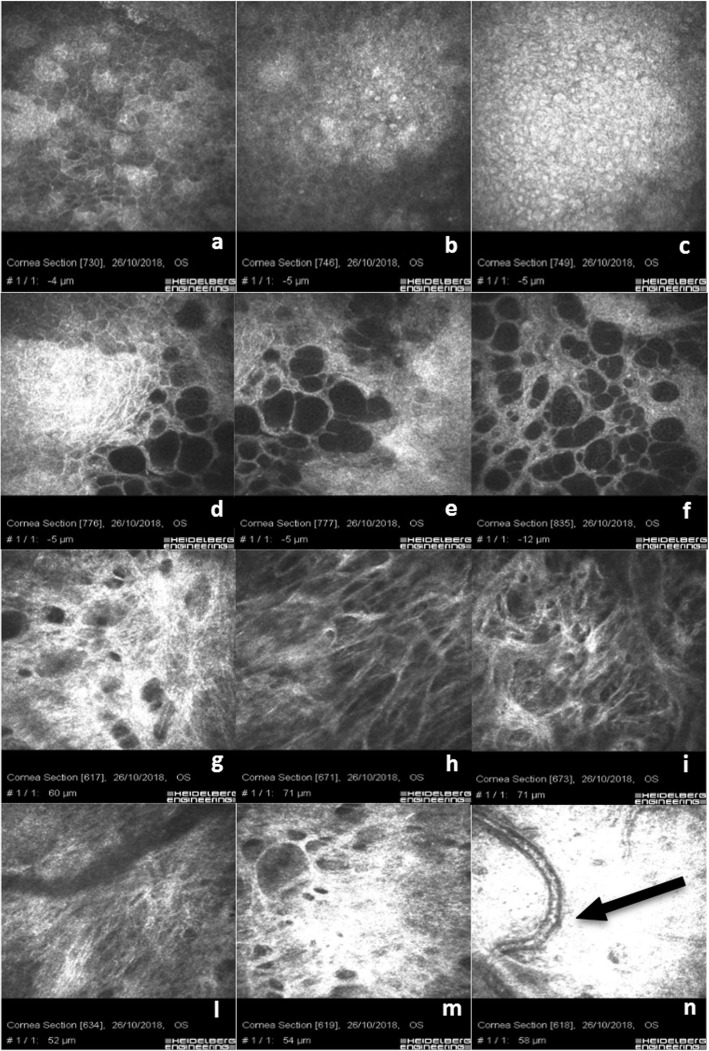
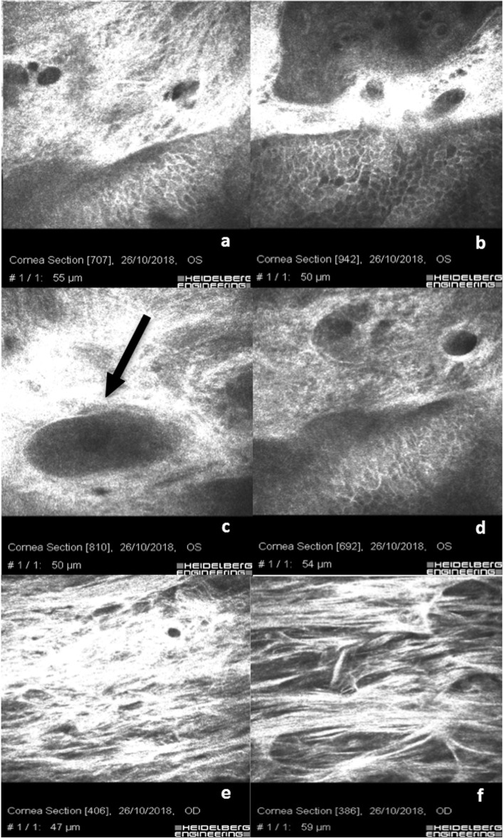
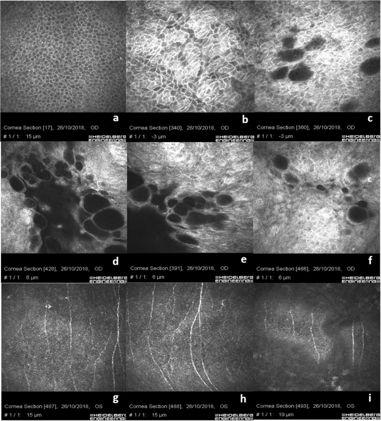
Results: The Xen 45 Gel stent, if properly positioned in the sub-conjunctival space preserves goblet cells and limits ocular surface inflammation. Regular corneal epithelial cells with micro-cysts, and normo-reflective sub-epithelial nerve plexus are documented by IVCM. In sub Tenon's implants an alternative lamellar intra-scleral filtration is detectable. Combined surgical procedures show a noticeable number of inflammatory cells with rare micro-cysts. Post-trabeculectomy inflammatory reaction is more evident than Xen 45 Gel Stent associated surgical procedures, but less than medical therapy where a conspicuous presence of Langerhans cells, peri-neural infiltrates, marked loss of goblet cells and fibrosis is visible.
Conclusion: Ocular surface inflammation was more notable in topical therapy than after trabeculectomy, which itself causes more inflammation than XEN Gel stents.

 求助内容:
求助内容: 应助结果提醒方式:


