Matteo Gravina, Grazia Casavecchia, Vincenzo Manuppelli, Antonio Totaro, Luca Macarini, Matteo Di Biase, Natale Daniele Brunetti
{"title":"二尖瓣环形钙化:CMR能识别干酪样坏死吗?","authors":"Matteo Gravina, Grazia Casavecchia, Vincenzo Manuppelli, Antonio Totaro, Luca Macarini, Matteo Di Biase, Natale Daniele Brunetti","doi":"10.1556/1646.10.2018.47","DOIUrl":null,"url":null,"abstract":"<p><p>Mitral annular calcification (MAC) can resemble an intracardiac mass and it is defined as a chronic degeneration of the mitral annulus. Often reported is caseous mitral annulus calcification (CMAC), a periannular, extensive calcification resembling a tumor. We report the case of a 68-year-old woman who had been hospitalized for palpitations and dyspnea. The transthoracic and transesophageal echocardiography revealed a non-homogeneous, slightly mobile, round mass, attached to the ventricular side of posterior mitral leaflet, with central echo-lucent area and without acoustic shadowing. Therefore, a cardiac magnetic resonance (CMR) was performed; delayed enhancement sequences showed a non-enhanced central core surrounded by a hyperenhanced rim (fibrous cap). To confirm the diagnosis, a multidetector computed tomography (MDCT) was performed; the MDCT showed a hyperdense mass with a hypodense center and a calcified peripheral rim. The central content had heterogeneous fluid density without significant contrast enhancement. The MDCT findings were considered highly suggestive of CMAC. CMR may be useful for the identification and definition of pericardial and myocardial masses and CMAC.</p>","PeriodicalId":45181,"journal":{"name":"Interventional Medicine and Applied Science","volume":"11 1","pages":"71-73"},"PeriodicalIF":0.0000,"publicationDate":"2019-03-01","publicationTypes":"Journal Article","fieldsOfStudy":null,"isOpenAccess":false,"openAccessPdf":"https://sci-hub-pdf.com/10.1556/1646.10.2018.47","citationCount":"1","resultStr":"{\"title\":\"Mitral annular calcification: Can CMR be useful in identifying caseous necrosis?\",\"authors\":\"Matteo Gravina, Grazia Casavecchia, Vincenzo Manuppelli, Antonio Totaro, Luca Macarini, Matteo Di Biase, Natale Daniele Brunetti\",\"doi\":\"10.1556/1646.10.2018.47\",\"DOIUrl\":null,\"url\":null,\"abstract\":\"<p><p>Mitral annular calcification (MAC) can resemble an intracardiac mass and it is defined as a chronic degeneration of the mitral annulus. Often reported is caseous mitral annulus calcification (CMAC), a periannular, extensive calcification resembling a tumor. We report the case of a 68-year-old woman who had been hospitalized for palpitations and dyspnea. The transthoracic and transesophageal echocardiography revealed a non-homogeneous, slightly mobile, round mass, attached to the ventricular side of posterior mitral leaflet, with central echo-lucent area and without acoustic shadowing. Therefore, a cardiac magnetic resonance (CMR) was performed; delayed enhancement sequences showed a non-enhanced central core surrounded by a hyperenhanced rim (fibrous cap). To confirm the diagnosis, a multidetector computed tomography (MDCT) was performed; the MDCT showed a hyperdense mass with a hypodense center and a calcified peripheral rim. The central content had heterogeneous fluid density without significant contrast enhancement. The MDCT findings were considered highly suggestive of CMAC. CMR may be useful for the identification and definition of pericardial and myocardial masses and CMAC.</p>\",\"PeriodicalId\":45181,\"journal\":{\"name\":\"Interventional Medicine and Applied Science\",\"volume\":\"11 1\",\"pages\":\"71-73\"},\"PeriodicalIF\":0.0000,\"publicationDate\":\"2019-03-01\",\"publicationTypes\":\"Journal Article\",\"fieldsOfStudy\":null,\"isOpenAccess\":false,\"openAccessPdf\":\"https://sci-hub-pdf.com/10.1556/1646.10.2018.47\",\"citationCount\":\"1\",\"resultStr\":null,\"platform\":\"Semanticscholar\",\"paperid\":null,\"PeriodicalName\":\"Interventional Medicine and Applied Science\",\"FirstCategoryId\":\"1085\",\"ListUrlMain\":\"https://doi.org/10.1556/1646.10.2018.47\",\"RegionNum\":0,\"RegionCategory\":null,\"ArticlePicture\":[],\"TitleCN\":null,\"AbstractTextCN\":null,\"PMCID\":null,\"EPubDate\":\"\",\"PubModel\":\"\",\"JCR\":\"Q2\",\"JCRName\":\"Medicine\",\"Score\":null,\"Total\":0}","platform":"Semanticscholar","paperid":null,"PeriodicalName":"Interventional Medicine and Applied Science","FirstCategoryId":"1085","ListUrlMain":"https://doi.org/10.1556/1646.10.2018.47","RegionNum":0,"RegionCategory":null,"ArticlePicture":[],"TitleCN":null,"AbstractTextCN":null,"PMCID":null,"EPubDate":"","PubModel":"","JCR":"Q2","JCRName":"Medicine","Score":null,"Total":0}
Mitral annular calcification: Can CMR be useful in identifying caseous necrosis?
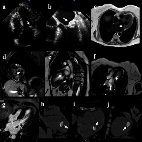
Mitral annular calcification (MAC) can resemble an intracardiac mass and it is defined as a chronic degeneration of the mitral annulus. Often reported is caseous mitral annulus calcification (CMAC), a periannular, extensive calcification resembling a tumor. We report the case of a 68-year-old woman who had been hospitalized for palpitations and dyspnea. The transthoracic and transesophageal echocardiography revealed a non-homogeneous, slightly mobile, round mass, attached to the ventricular side of posterior mitral leaflet, with central echo-lucent area and without acoustic shadowing. Therefore, a cardiac magnetic resonance (CMR) was performed; delayed enhancement sequences showed a non-enhanced central core surrounded by a hyperenhanced rim (fibrous cap). To confirm the diagnosis, a multidetector computed tomography (MDCT) was performed; the MDCT showed a hyperdense mass with a hypodense center and a calcified peripheral rim. The central content had heterogeneous fluid density without significant contrast enhancement. The MDCT findings were considered highly suggestive of CMAC. CMR may be useful for the identification and definition of pericardial and myocardial masses and CMAC.

 求助内容:
求助内容: 应助结果提醒方式:


