Macit Kalçık, Mahmut Yesin, Ahmet Güner, Emrah Bayam, Mucahit Yetim, Tolga Doğan, Lütfü Bekar, Oğuzhan Çelik, Yusuf Karavelioğlu
{"title":"微血管性心绞痛患者心外膜脂肪组织厚度的超声心动图测量。","authors":"Macit Kalçık, Mahmut Yesin, Ahmet Güner, Emrah Bayam, Mucahit Yetim, Tolga Doğan, Lütfü Bekar, Oğuzhan Çelik, Yusuf Karavelioğlu","doi":"10.1556/1646.11.2019.12","DOIUrl":null,"url":null,"abstract":"<p><strong>Introduction: </strong>Impaired coronary microcirculation, inflammation, and endothelial dysfunction were reported etiological factors for microvascular angina (MVA). Recently, increased epicardial adipose tissue (EAT) thickness has been associated with hypertension, metabolic syndrome, and coronary artery disease in general population. In this study, we aimed to evaluate the EAT thickness in patients with MVA.</p><p><strong>Methods: </strong>This study enrolled 200 patients (83 males; mean age: 55.4 ± 8.2 years) who have been diagnosed with MVA and 200 controls (89 males; mean age: 54.4 ± 8.5 years). All patients underwent transthoracic echocardiography, and EAT thickness was measured from a parasternal long-axis view as the hypoechoic space on the right ventricular free wall.</p><p><strong>Results: </strong>The mean EAT thickness was significantly higher in MVA patients than the controls (5.5 ± 1.1 vs. 4.9 ± 0.7 mm; <i>p</i> < 0.001). Multiple logistic regression analysis showed that increased EAT thickness was an independent predictor of MVA (OR = 1.183, 95% CI = 1.063-1.489; <i>p</i> = 0.023). In receiver operating characteristic curve analyses, EAT thickness above 5.3 mm predicted MVA with a sentivity of 68% and a specificity of 63% (AUC = 0.711, 95% CI = 0.659-0.762; <i>p</i> < 0.001).</p><p><strong>Conclusions: </strong>The EAT thickness was observed significantly higher in MVA patients as compared to controls. Increased EAT thickness may be associated with mechanisms that play a major role in the pathogenesis of MVA.</p>","PeriodicalId":45181,"journal":{"name":"Interventional Medicine and Applied Science","volume":"11 2","pages":"106-111"},"PeriodicalIF":0.0000,"publicationDate":"2019-06-01","publicationTypes":"Journal Article","fieldsOfStudy":null,"isOpenAccess":false,"openAccessPdf":"https://sci-hub-pdf.com/10.1556/1646.11.2019.12","citationCount":"0","resultStr":"{\"title\":\"Echocardiographic measurement of epicardial adipose tissue thickness in patients with microvascular angina.\",\"authors\":\"Macit Kalçık, Mahmut Yesin, Ahmet Güner, Emrah Bayam, Mucahit Yetim, Tolga Doğan, Lütfü Bekar, Oğuzhan Çelik, Yusuf Karavelioğlu\",\"doi\":\"10.1556/1646.11.2019.12\",\"DOIUrl\":null,\"url\":null,\"abstract\":\"<p><strong>Introduction: </strong>Impaired coronary microcirculation, inflammation, and endothelial dysfunction were reported etiological factors for microvascular angina (MVA). Recently, increased epicardial adipose tissue (EAT) thickness has been associated with hypertension, metabolic syndrome, and coronary artery disease in general population. In this study, we aimed to evaluate the EAT thickness in patients with MVA.</p><p><strong>Methods: </strong>This study enrolled 200 patients (83 males; mean age: 55.4 ± 8.2 years) who have been diagnosed with MVA and 200 controls (89 males; mean age: 54.4 ± 8.5 years). All patients underwent transthoracic echocardiography, and EAT thickness was measured from a parasternal long-axis view as the hypoechoic space on the right ventricular free wall.</p><p><strong>Results: </strong>The mean EAT thickness was significantly higher in MVA patients than the controls (5.5 ± 1.1 vs. 4.9 ± 0.7 mm; <i>p</i> < 0.001). Multiple logistic regression analysis showed that increased EAT thickness was an independent predictor of MVA (OR = 1.183, 95% CI = 1.063-1.489; <i>p</i> = 0.023). In receiver operating characteristic curve analyses, EAT thickness above 5.3 mm predicted MVA with a sentivity of 68% and a specificity of 63% (AUC = 0.711, 95% CI = 0.659-0.762; <i>p</i> < 0.001).</p><p><strong>Conclusions: </strong>The EAT thickness was observed significantly higher in MVA patients as compared to controls. Increased EAT thickness may be associated with mechanisms that play a major role in the pathogenesis of MVA.</p>\",\"PeriodicalId\":45181,\"journal\":{\"name\":\"Interventional Medicine and Applied Science\",\"volume\":\"11 2\",\"pages\":\"106-111\"},\"PeriodicalIF\":0.0000,\"publicationDate\":\"2019-06-01\",\"publicationTypes\":\"Journal Article\",\"fieldsOfStudy\":null,\"isOpenAccess\":false,\"openAccessPdf\":\"https://sci-hub-pdf.com/10.1556/1646.11.2019.12\",\"citationCount\":\"0\",\"resultStr\":null,\"platform\":\"Semanticscholar\",\"paperid\":null,\"PeriodicalName\":\"Interventional Medicine and Applied Science\",\"FirstCategoryId\":\"1085\",\"ListUrlMain\":\"https://doi.org/10.1556/1646.11.2019.12\",\"RegionNum\":0,\"RegionCategory\":null,\"ArticlePicture\":[],\"TitleCN\":null,\"AbstractTextCN\":null,\"PMCID\":null,\"EPubDate\":\"\",\"PubModel\":\"\",\"JCR\":\"Q2\",\"JCRName\":\"Medicine\",\"Score\":null,\"Total\":0}","platform":"Semanticscholar","paperid":null,"PeriodicalName":"Interventional Medicine and Applied Science","FirstCategoryId":"1085","ListUrlMain":"https://doi.org/10.1556/1646.11.2019.12","RegionNum":0,"RegionCategory":null,"ArticlePicture":[],"TitleCN":null,"AbstractTextCN":null,"PMCID":null,"EPubDate":"","PubModel":"","JCR":"Q2","JCRName":"Medicine","Score":null,"Total":0}
引用次数: 0
摘要
导语:冠状动脉微循环受损、炎症和内皮功能障碍是微血管心绞痛(MVA)的病因。最近,在一般人群中,心外膜脂肪组织(EAT)厚度增加与高血压、代谢综合征和冠状动脉疾病有关。在本研究中,我们旨在评估MVA患者的EAT厚度。方法:本研究纳入200例患者(男性83例;平均年龄:55.4±8.2岁),对照组200例(男性89例;平均年龄:54.4±8.5岁)。所有患者都进行了经胸超声心动图检查,并从胸骨旁长轴视图作为右心室游离壁的低回声空间测量EAT厚度。结果:MVA患者的平均EAT厚度显著高于对照组(5.5±1.1 vs. 4.9±0.7 mm;p = 0.023)。在受试者工作特征曲线分析中,EAT厚度大于5.3 mm预测MVA的敏感性为68%,特异性为63% (AUC = 0.711, 95% CI = 0.659-0.762;结论:与对照组相比,MVA患者的EAT厚度明显增高。EAT厚度增加可能与在MVA发病机制中起主要作用的机制有关。
Echocardiographic measurement of epicardial adipose tissue thickness in patients with microvascular angina.
Introduction: Impaired coronary microcirculation, inflammation, and endothelial dysfunction were reported etiological factors for microvascular angina (MVA). Recently, increased epicardial adipose tissue (EAT) thickness has been associated with hypertension, metabolic syndrome, and coronary artery disease in general population. In this study, we aimed to evaluate the EAT thickness in patients with MVA.
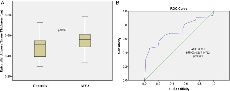
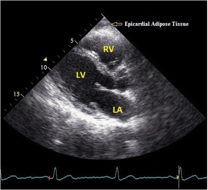
Methods: This study enrolled 200 patients (83 males; mean age: 55.4 ± 8.2 years) who have been diagnosed with MVA and 200 controls (89 males; mean age: 54.4 ± 8.5 years). All patients underwent transthoracic echocardiography, and EAT thickness was measured from a parasternal long-axis view as the hypoechoic space on the right ventricular free wall.
Results: The mean EAT thickness was significantly higher in MVA patients than the controls (5.5 ± 1.1 vs. 4.9 ± 0.7 mm; p < 0.001). Multiple logistic regression analysis showed that increased EAT thickness was an independent predictor of MVA (OR = 1.183, 95% CI = 1.063-1.489; p = 0.023). In receiver operating characteristic curve analyses, EAT thickness above 5.3 mm predicted MVA with a sentivity of 68% and a specificity of 63% (AUC = 0.711, 95% CI = 0.659-0.762; p < 0.001).
Conclusions: The EAT thickness was observed significantly higher in MVA patients as compared to controls. Increased EAT thickness may be associated with mechanisms that play a major role in the pathogenesis of MVA.

 求助内容:
求助内容: 应助结果提醒方式:


