{"title":"冠状动脉慢血流患者同型半胱氨酸和非对称二甲基精氨酸水平的评价。","authors":"Erkan Demirci, Oğuzhan Çelik, Macit Kalçık, Lütfü Bekar, Mucahit Yetim, Tolga Doğan","doi":"10.1556/1646.11.2019.07","DOIUrl":null,"url":null,"abstract":"<p><strong>Background: </strong>Previous studies have demonstrated that homocysteine and asymmetric dimethyl arginine (ADMA) levels were strongly associated with cardiovascular diseases including coronary artery disease. The aim of this study was to investigate the role of plasma homocysteine and ADMA levels in the pathogenesis of coronary slow flow (CSF) phenomenon.</p><p><strong>Methods: </strong>Twenty-three patients with CSF and 25 controls with normal coronary flow were included in this study. The quantitative measurement of coronary blood flow was performed using the thrombolysis in myocardial infarction frame count method. Plasma homocysteine and ADMA levels were determined using enzymatic assays from venous blood samples.</p><p><strong>Results: </strong>The patients with CSF had significantly higher plasma homocysteine levels than controls (16.2 ± 7.6 vs. 12.2 ± 2.2 μM/L; <i>p</i> = 0.023). The uric acid levels were significantly higher in CSF group than controls (5.4 ± 1.1 vs. 4.6 ± 0.9 mg/dl; <i>p</i> = 0.011). Plasma ADMA levels were also higher in the CSF group; however, this was not statistically significant (0.6 ± 0.1 vs. 0.5 ± 0.2 μM/L; <i>p</i> = 0.475).</p><p><strong>Conclusions: </strong>Increased homocysteine and uric acid levels may play an important role in the pathogenesis of CSF. Further large scale studies are required to determine the relationship between ADMA levels and CSF.</p>","PeriodicalId":45181,"journal":{"name":"Interventional Medicine and Applied Science","volume":"11 2","pages":"89-94"},"PeriodicalIF":0.0000,"publicationDate":"2019-06-01","publicationTypes":"Journal Article","fieldsOfStudy":null,"isOpenAccess":false,"openAccessPdf":"https://sci-hub-pdf.com/10.1556/1646.11.2019.07","citationCount":"9","resultStr":"{\"title\":\"Evaluation of homocystein and asymmetric dimethyl arginine levels in patients with coronary slow flow phenomenon.\",\"authors\":\"Erkan Demirci, Oğuzhan Çelik, Macit Kalçık, Lütfü Bekar, Mucahit Yetim, Tolga Doğan\",\"doi\":\"10.1556/1646.11.2019.07\",\"DOIUrl\":null,\"url\":null,\"abstract\":\"<p><strong>Background: </strong>Previous studies have demonstrated that homocysteine and asymmetric dimethyl arginine (ADMA) levels were strongly associated with cardiovascular diseases including coronary artery disease. The aim of this study was to investigate the role of plasma homocysteine and ADMA levels in the pathogenesis of coronary slow flow (CSF) phenomenon.</p><p><strong>Methods: </strong>Twenty-three patients with CSF and 25 controls with normal coronary flow were included in this study. The quantitative measurement of coronary blood flow was performed using the thrombolysis in myocardial infarction frame count method. Plasma homocysteine and ADMA levels were determined using enzymatic assays from venous blood samples.</p><p><strong>Results: </strong>The patients with CSF had significantly higher plasma homocysteine levels than controls (16.2 ± 7.6 vs. 12.2 ± 2.2 μM/L; <i>p</i> = 0.023). The uric acid levels were significantly higher in CSF group than controls (5.4 ± 1.1 vs. 4.6 ± 0.9 mg/dl; <i>p</i> = 0.011). Plasma ADMA levels were also higher in the CSF group; however, this was not statistically significant (0.6 ± 0.1 vs. 0.5 ± 0.2 μM/L; <i>p</i> = 0.475).</p><p><strong>Conclusions: </strong>Increased homocysteine and uric acid levels may play an important role in the pathogenesis of CSF. Further large scale studies are required to determine the relationship between ADMA levels and CSF.</p>\",\"PeriodicalId\":45181,\"journal\":{\"name\":\"Interventional Medicine and Applied Science\",\"volume\":\"11 2\",\"pages\":\"89-94\"},\"PeriodicalIF\":0.0000,\"publicationDate\":\"2019-06-01\",\"publicationTypes\":\"Journal Article\",\"fieldsOfStudy\":null,\"isOpenAccess\":false,\"openAccessPdf\":\"https://sci-hub-pdf.com/10.1556/1646.11.2019.07\",\"citationCount\":\"9\",\"resultStr\":null,\"platform\":\"Semanticscholar\",\"paperid\":null,\"PeriodicalName\":\"Interventional Medicine and Applied Science\",\"FirstCategoryId\":\"1085\",\"ListUrlMain\":\"https://doi.org/10.1556/1646.11.2019.07\",\"RegionNum\":0,\"RegionCategory\":null,\"ArticlePicture\":[],\"TitleCN\":null,\"AbstractTextCN\":null,\"PMCID\":null,\"EPubDate\":\"\",\"PubModel\":\"\",\"JCR\":\"Q2\",\"JCRName\":\"Medicine\",\"Score\":null,\"Total\":0}","platform":"Semanticscholar","paperid":null,"PeriodicalName":"Interventional Medicine and Applied Science","FirstCategoryId":"1085","ListUrlMain":"https://doi.org/10.1556/1646.11.2019.07","RegionNum":0,"RegionCategory":null,"ArticlePicture":[],"TitleCN":null,"AbstractTextCN":null,"PMCID":null,"EPubDate":"","PubModel":"","JCR":"Q2","JCRName":"Medicine","Score":null,"Total":0}
引用次数: 9
摘要
背景:以往的研究表明,同型半胱氨酸和不对称二甲基精氨酸(ADMA)水平与包括冠状动脉疾病在内的心血管疾病密切相关。本研究旨在探讨血浆同型半胱氨酸和ADMA水平在冠状动脉慢流(CSF)发病机制中的作用。方法:23例脑脊液患者和25例冠状动脉血流正常的对照组。采用心肌梗死溶栓框架计数法定量测定冠状动脉血流量。血浆同型半胱氨酸和ADMA水平用静脉血样品的酶法测定。结果:脑脊液患者血浆同型半胱氨酸水平显著高于对照组(16.2±7.6 vs 12.2±2.2 μM/L;p = 0.023)。脑脊液组尿酸水平显著高于对照组(5.4±1.1 vs. 4.6±0.9 mg/dl;p = 0.011)。脑脊液组血浆ADMA水平也较高;但差异无统计学意义(0.6±0.1 vs. 0.5±0.2 μM/L;p = 0.475)。结论:同型半胱氨酸和尿酸水平升高可能在脑脊液的发病机制中起重要作用。需要进一步的大规模研究来确定ADMA水平与CSF之间的关系。
Evaluation of homocystein and asymmetric dimethyl arginine levels in patients with coronary slow flow phenomenon.
Background: Previous studies have demonstrated that homocysteine and asymmetric dimethyl arginine (ADMA) levels were strongly associated with cardiovascular diseases including coronary artery disease. The aim of this study was to investigate the role of plasma homocysteine and ADMA levels in the pathogenesis of coronary slow flow (CSF) phenomenon.
Methods: Twenty-three patients with CSF and 25 controls with normal coronary flow were included in this study. The quantitative measurement of coronary blood flow was performed using the thrombolysis in myocardial infarction frame count method. Plasma homocysteine and ADMA levels were determined using enzymatic assays from venous blood samples.
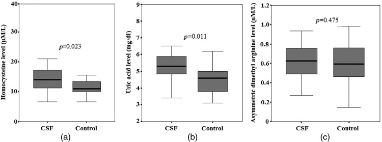
Results: The patients with CSF had significantly higher plasma homocysteine levels than controls (16.2 ± 7.6 vs. 12.2 ± 2.2 μM/L; p = 0.023). The uric acid levels were significantly higher in CSF group than controls (5.4 ± 1.1 vs. 4.6 ± 0.9 mg/dl; p = 0.011). Plasma ADMA levels were also higher in the CSF group; however, this was not statistically significant (0.6 ± 0.1 vs. 0.5 ± 0.2 μM/L; p = 0.475).
Conclusions: Increased homocysteine and uric acid levels may play an important role in the pathogenesis of CSF. Further large scale studies are required to determine the relationship between ADMA levels and CSF.

 求助内容:
求助内容: 应助结果提醒方式:


