{"title":"Mirizzi综合征合并胆总管瘘:从无症状胆囊结石发展到手术的观察。","authors":"Hiroyuki Sugo, Yuuki Sekine, Naoki Iwanaga, Shigefumi Neshime, Michio Machida","doi":"10.1155/2020/2049525","DOIUrl":null,"url":null,"abstract":"<p><p>Despite a considerable number of reports of Mirizzi syndrome, none have described the process of its development from simple cholecystolithiasis. We report an extremely rare case of Mirizzi syndrome in which it was possible to observe the process of development of cholecystobiliary fistula from asymptomatic cholecystolithiasis until unavoidable surgical intervention 4 years later. A 68-year-old woman presented at our hospital with right upper quadrant pain. She had been diagnosed as having asymptomatic cholecystolithiasis 4 years previously. Diagnostic abdominal computed tomography (CT) had revealed a 1.9 cm radiopaque stone, and thereafter, the patient had been monitored by imaging alone. CT conducted 6 months before the present admission revealed that the gallbladder stone was compressing the common hepatic duct, although the patient remained asymptomatic. On admission, abdominal CT showed that the gallbladder stone was obstructing the common bile duct with dilatation of the intrahepatic duct. Endoscopic retrograde cholangiopancreatography revealed a round filling defect at the confluence of the common bile duct and the image of the cystic duct; therefore, the patient was categorized as having Mirizzi syndrome type III, according to the Csendes classification. Intraoperative findings revealed a cholecystobiliary fistula involving up to two-thirds of the circumference of the common bile duct.</p>","PeriodicalId":30326,"journal":{"name":"Case Reports in Radiology","volume":"2020 ","pages":"2049525"},"PeriodicalIF":0.0000,"publicationDate":"2020-01-27","publicationTypes":"Journal Article","fieldsOfStudy":null,"isOpenAccess":false,"openAccessPdf":"https://sci-hub-pdf.com/10.1155/2020/2049525","citationCount":"0","resultStr":"{\"title\":\"Mirizzi Syndrome with Cholecystobiliary Fistula: Observation of Development from Asymptomatic Cholecystolithiasis to Surgery.\",\"authors\":\"Hiroyuki Sugo, Yuuki Sekine, Naoki Iwanaga, Shigefumi Neshime, Michio Machida\",\"doi\":\"10.1155/2020/2049525\",\"DOIUrl\":null,\"url\":null,\"abstract\":\"<p><p>Despite a considerable number of reports of Mirizzi syndrome, none have described the process of its development from simple cholecystolithiasis. We report an extremely rare case of Mirizzi syndrome in which it was possible to observe the process of development of cholecystobiliary fistula from asymptomatic cholecystolithiasis until unavoidable surgical intervention 4 years later. A 68-year-old woman presented at our hospital with right upper quadrant pain. She had been diagnosed as having asymptomatic cholecystolithiasis 4 years previously. Diagnostic abdominal computed tomography (CT) had revealed a 1.9 cm radiopaque stone, and thereafter, the patient had been monitored by imaging alone. CT conducted 6 months before the present admission revealed that the gallbladder stone was compressing the common hepatic duct, although the patient remained asymptomatic. On admission, abdominal CT showed that the gallbladder stone was obstructing the common bile duct with dilatation of the intrahepatic duct. Endoscopic retrograde cholangiopancreatography revealed a round filling defect at the confluence of the common bile duct and the image of the cystic duct; therefore, the patient was categorized as having Mirizzi syndrome type III, according to the Csendes classification. Intraoperative findings revealed a cholecystobiliary fistula involving up to two-thirds of the circumference of the common bile duct.</p>\",\"PeriodicalId\":30326,\"journal\":{\"name\":\"Case Reports in Radiology\",\"volume\":\"2020 \",\"pages\":\"2049525\"},\"PeriodicalIF\":0.0000,\"publicationDate\":\"2020-01-27\",\"publicationTypes\":\"Journal Article\",\"fieldsOfStudy\":null,\"isOpenAccess\":false,\"openAccessPdf\":\"https://sci-hub-pdf.com/10.1155/2020/2049525\",\"citationCount\":\"0\",\"resultStr\":null,\"platform\":\"Semanticscholar\",\"paperid\":null,\"PeriodicalName\":\"Case Reports in Radiology\",\"FirstCategoryId\":\"1085\",\"ListUrlMain\":\"https://doi.org/10.1155/2020/2049525\",\"RegionNum\":0,\"RegionCategory\":null,\"ArticlePicture\":[],\"TitleCN\":null,\"AbstractTextCN\":null,\"PMCID\":null,\"EPubDate\":\"2020/1/1 0:00:00\",\"PubModel\":\"eCollection\",\"JCR\":\"\",\"JCRName\":\"\",\"Score\":null,\"Total\":0}","platform":"Semanticscholar","paperid":null,"PeriodicalName":"Case Reports in Radiology","FirstCategoryId":"1085","ListUrlMain":"https://doi.org/10.1155/2020/2049525","RegionNum":0,"RegionCategory":null,"ArticlePicture":[],"TitleCN":null,"AbstractTextCN":null,"PMCID":null,"EPubDate":"2020/1/1 0:00:00","PubModel":"eCollection","JCR":"","JCRName":"","Score":null,"Total":0}
Mirizzi Syndrome with Cholecystobiliary Fistula: Observation of Development from Asymptomatic Cholecystolithiasis to Surgery.
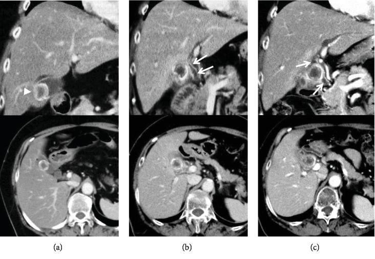
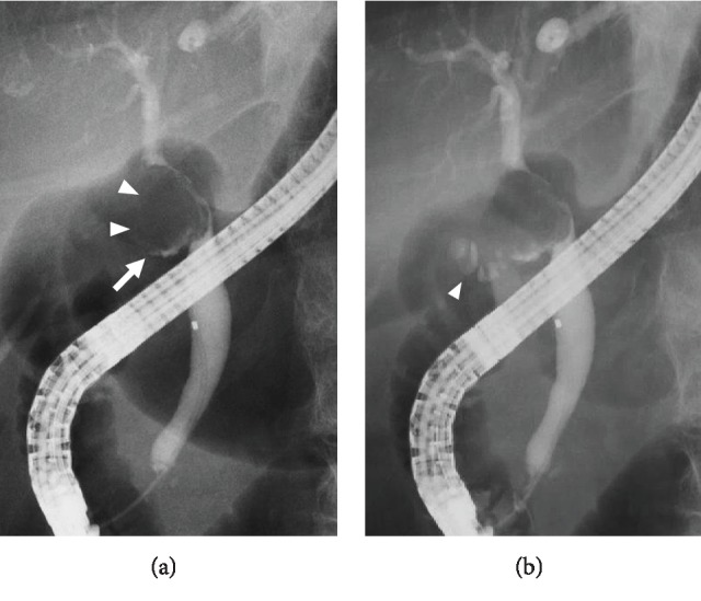
Despite a considerable number of reports of Mirizzi syndrome, none have described the process of its development from simple cholecystolithiasis. We report an extremely rare case of Mirizzi syndrome in which it was possible to observe the process of development of cholecystobiliary fistula from asymptomatic cholecystolithiasis until unavoidable surgical intervention 4 years later. A 68-year-old woman presented at our hospital with right upper quadrant pain. She had been diagnosed as having asymptomatic cholecystolithiasis 4 years previously. Diagnostic abdominal computed tomography (CT) had revealed a 1.9 cm radiopaque stone, and thereafter, the patient had been monitored by imaging alone. CT conducted 6 months before the present admission revealed that the gallbladder stone was compressing the common hepatic duct, although the patient remained asymptomatic. On admission, abdominal CT showed that the gallbladder stone was obstructing the common bile duct with dilatation of the intrahepatic duct. Endoscopic retrograde cholangiopancreatography revealed a round filling defect at the confluence of the common bile duct and the image of the cystic duct; therefore, the patient was categorized as having Mirizzi syndrome type III, according to the Csendes classification. Intraoperative findings revealed a cholecystobiliary fistula involving up to two-thirds of the circumference of the common bile duct.


 求助内容:
求助内容: 应助结果提醒方式:


